若您需要咨询产品或有任何技术问题,请通过官方电话 400 885 9050 或邮箱 info.cn@stemcell.com 与我们联系。

MesenCult™-ACF软骨细胞分化试剂盒

用于MSCs向软骨细胞分化的无动物成分培养基
只有 %1
¥3,050.00

产品号 #(选择产品)

产品号 #05455_C

用于MSCs向软骨细胞分化的无动物成分培养基

产品优势

  • 无动物成分(ACF)配方
  • 最快需要14天,实现最少3 × 10^5 MSCs高效地软骨分化
  • 软骨相关转录因子Acan、Col2a、Sox9和Col10a的强表达;肥大相关转录因子Mmp13低表达
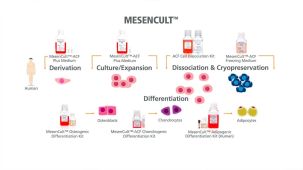
  • 优化的完整无动物成分(ACF)产品线,用于MSC的分离、扩增、冻存和软骨分化

产品组分包括

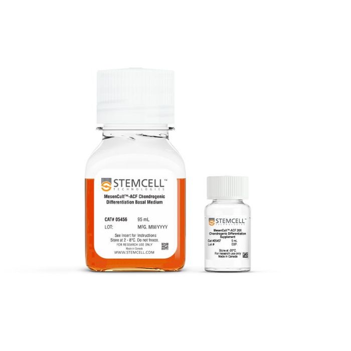
  • MesenCult™-ACF软骨分化基础培养基,95 mL
  • MesenCult™-ACF 20X 软骨分化补充剂, 5 mL
专为您的实验方案打造的产品
要查看实验方案所需的所有配套产品,请参阅《实验方案与技术文档》

总览

使用MesenCult™-ACF软骨分化试剂盒,从人间充质基质细胞(MSCs)和hPSC衍生的间充质祖细胞中生成软骨谱系细胞(包括软骨细胞)。这种完全无动物成分(ACF)的配方可从低至3×10⁵个细胞开始实现高效的软骨生成分化和基质沉积。生成的软骨细胞最早在第14天即可检测到软骨生成。该培养基与源自骨髓、脂肪组织或滑膜的MSC兼容,并适用于在含血清或ACF条件下扩增的细胞。

将此试剂盒作为完整的MSC功能评估工作流程的一部分使用。与MesenCult™脂肪生成分化试剂盒(人)MesenCult™成骨分化试剂盒(人)配合使用,以评估三谱系分化潜能并验证MSC的身份和功能。这些即用型试剂能够可靠地评估MSC的身份和功能,同时确保在整个MesenCult™工作流程中获得可重复的谱系特异性结果。


分类
专用培养基
 
细胞类型
软骨细胞,间充质干/祖细胞
 
种属

 
应用
细胞培养,分化
 
品牌
MesenCult
 
研究领域
药物发现和毒性检测,干细胞生物学
 
制剂类别
不含动物成分,无血清
 

实验数据

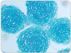
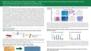
MesenCult™-ACF Chondrogenic Differentiation Medium Induces Robust Chondrogenic Differentiation of Human MSCs

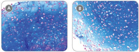
Figure 1. MesenCult™-ACF Chondrogenic Differentiation Medium Induces Robust Chondrogenic Differentiation of Human MSCs

Human BM-derived MSCs were cultured in MesenCult™-ACF Medium then differentiated to the chondrogenic lineage using MesenCult™-ACF Chondrogenic Differentiation Medium. Robust chondrogenic differentiation was observed (A) starting with as few as 3 x 10⁵ MSCs, or (B) when differentiating for just 14 days starting with 5 x 10⁵ MSCs.

Chondrogenic Differentiation of Human MSCs Is More Robust With Fewer Hypertrophic Chondrocytes Using MesenCult™-ACF Chondrogenic Differentiation Medium Compared to Competitor Media

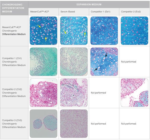
Figure 2. Chondrogenic Differentiation of Human MSCs Is More Robust With Fewer Hypertrophic Chondrocytes Using MesenCult™-ACF Chondrogenic Differentiation Medium Compared to Competitor Media

Human BM-derived MSCs culture-expanded for up to two passages in MesenCult™-ACF Medium, serum-based medium, or one of two commercially available media (Competitor 1 (Ex1) and Competitor 2 (Ex2)), were then differentiated to the chondrogenic lineage starting with 5 x 10⁵ MSCs and either using MesenCult™-ACF Chondrogenic Differentiation Medium or one of several commercially available chondrogenic differentiation media (Ch1, Ch2 or Ch3) for 21 days. More robust and uniform chondrogenic differentiation was observed when the MSCs were differentiated in MesenCult™-ACF Chondrogenic Differentiation Medium compared to the other commercially available chondrogenic differentiation media (Ch1, Ch2 and Ch3), irrespective of the expansion medium used to culture the MSCs prior to differentiation. Cultures differentiated using MesenCult™-ACF Chondrogenic Differentiation Medium displayed an abundance of isogenous groups (yellow arrows), suggesting there is proliferation of differentiating chondrocyte progenitors. Few hypertrophic chondrocytes (black arrows) are seen in cultures differentiated with MesenCult™-ACF Chondrogenic Differentiation Medium, suggesting the maintenance of chondrogenic activity throughout the culturing period.

MesenCult™-ACF Chondrogenic Differentiation Medium Induces Stronger and More Sustained Chondrogenic Transcript Expression Compared to Competitor Media

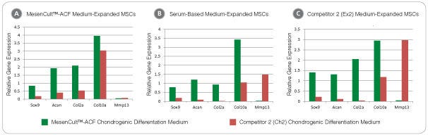
Figure 3. MesenCult™-ACF Chondrogenic Differentiation Medium Induces Stronger and More Sustained Chondrogenic Transcript Expression Compared to Competitor Media

Human BM-derived MSCs expanded in (A) MesenCult™-ACF Medium, (B) a serum-based medium or (C) Competitor 2 (Ex2) medium, were differentiated for 21 days with MesenCult™-ACF Chondrogenic Differentiation Medium and Competitor 2 (Ch2) chondrogenic differentiation medium. Regardless of the expansion medium initially used to culture the MSCs, differentiation using MesenCult™-ACF Chondrogenic Differentiation Medium led to a substantial up-regulation of the chondrogenic transcripts compared to Ch2. In addition, expression of the terminally-differentiated hypertrophic transcript Mmp13 was higher for Ch2 differentiated cultures compared to cultures differentiated with MesenCult™-ACF Chondrogenic Differentiation Medium.

Mouse MSCs Cultured in MesenCult™-ACF Chondrogenic Differentiation Medium Differentiate to Chondrocytes

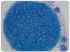
Figure 4. Mouse MSCs Cultured in MesenCult™-ACF Chondrogenic Differentiation Medium Differentiate to Chondrocytes

Mouse compact bone-derived MSCs were cultured using the MesenCult™ Proliferation Kit with MesenPure™ (Mouse, Catalog #05512) for 2 passages then differentiated by pellet culture with MesenCult™-ACF Chondrogenic Differentiation Medium for 21 days under normoxic (20% O2) conditions. Strong chondrogenic differentiation is indicated by dark-blue staining of the cartilage extracellular matrix and an abundance of isogenous chondrocyte groups.

Human MSC Chondrogenic Differentiation With AggreWell™800 Plates

Figure 5. Human MSC Chondrogenic Differentiation With AggreWell™800 Plates

Using centrifugation, 1 x 10^6 human MSCs were distributed evenly among 800 µm microwells in one well of an AggreWell™800 plate. Small aggregates of only ~3,300 cells per pellet were then differentiated to chondrocytes using MesenCult™-ACF Chondrogenic Differentiation Medium for 21 days under normoxic (20% O2) conditions.

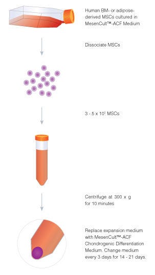
Procedure Overview: Human MSC Chondrogenic Differentiation Using MesenCult™-ACF Chondrogenic Differentiation Medium

Figure 6. Procedure Overview: Human MSC Chondrogenic Differentiation Using MesenCult™-ACF Chondrogenic Differentiation Medium

Aggregate culture is a useful method for inducing chondrogenic differentiation of human BM- and adipose-derived MSCs in a three-dimensional in vitro culture environment. MSCs are efficiently differentiated to the chondrogenic lineage using MesenCult™-ACF Chondrogenic Differentiation Medium in 14 - 21 days with 3 - 5 x 10⁵ cells.

产品说明书及文档

请在《产品说明书》中查找相关支持信息和使用说明,或浏览下方更多实验方案。

Document Type
Product Name
Catalog #
Lot #
Language
Catalog #
05455
Lot #
All
Language
English
Document Type
Safety Data Sheet 1
Catalog #
05455
Lot #
All
Language
English
Document Type
Safety Data Sheet 2
Catalog #
05455
Lot #
All
Language
English

应用领域

本产品专为以下研究领域设计,适用于工作流程中的高亮阶段。探索这些工作流程,了解更多我们为各研究领域提供的其他配套产品。

相关材料与文献

技术资料 (9)

文献 (14)

High-Efficiency Lentiviral Gene Modification of Primary Murine Bone-Marrow Mesenchymal Stem Cells. D. Gerace et al. Methods in molecular biology (Clifton,N.J.) 2019

Abstract

Lentiviral vectors are the method of choice for stable gene modification of a variety of cell types. However,the efficiency with which they transduce target cells varies significantly,in particular their typically poor capacity to transduce primary stem cells. Here we describe the isolation and enrichment of murine bone-marrow mesenchymal stem cells (MSCs) via fluorescence-activated cell sorting (FACS); the cloning,production,and concentration of high-titer second generation lentiviral vectors via combined tangential flow filtration (TFF) and ultracentrifugation; and the subsequent high-efficiency gene modification of MSCs into insulin-producing cells via overexpression of the furin-cleavable human insulin (INS-FUR) gene.
Promoting Osteogenic Differentiation of Human Adipose-Derived Stem Cells by Altering the Expression of Exosomal miRNA. S. Yang et al. Stem cells international 2019

Abstract

Human adipose-derived stem cells (ADSCs) can release exosomes; however,their specific functions remain elusive. In this study,we verified that exosomes derived from osteogenically differentiated ADSCs can promote osteogenic differentiation of ADSCs. Furthermore,in order to investigate the importance of exosomal microRNAs (miRNAs) in osteogenic differentiation of ADSCs,we used microarray assays to analyze the expression profiles of exosomal miRNAs derived from undifferentiated as well as osteogenically differentiated ADSCs; 201 miRNAs were upregulated and 33 miRNAs were downregulated between the two types of exosomes. Additionally,bioinformatic analyses,which included gene ontology analyses,pathway analysis,and miRNA-mRNA-network investigations,were performed. The results of these analyses revealed that the differentially expressed exosomal miRNAs participate in multiple biological processes,such as gene expression,synthesis of biomolecules,cell development,differentiation,and signal transduction,among others. Moreover,we found that these differentially expressed exosomal miRNAs connect osteogenic differentiation to processes such as axon guidance,MAPK signaling,and Wnt signaling. To the best of our knowledge,this is the first study to identify and characterize exosomal miRNAs derived from osteogenically differentiated ADSCs. This study confirms that alterations in the expression of exosomal miRNAs can promote osteogenic differentiation of ADSCs,which also provides the foundation for further research on the regulatory functions of exosomal miRNAs in the context of ADSC osteogenesis.
Mesenchymal stromal cells lower platelet activation and assist in platelet formation in vitro. A. Mendelson et al. JCI insight 2019 aug

Abstract

The complex process of platelet formation originates with the hematopoietic stem cell,which differentiates through the myeloid lineage,matures,and releases proplatelets into the BM sinusoids. How formed platelets maintain a low basal activation state in the circulation remains unknown. We identify Lepr+ stromal cells lining the BM sinusoids as important contributors to sustaining low platelet activation. Ablation of murine Lepr+ cells led to a decreased number of platelets in the circulation with an increased activation state. We developed a potentially novel culture system for supporting platelet formation in vitro using a unique population of CD51+PDGFRalpha+ perivascular cells,derived from human umbilical cord tissue,which display numerous mesenchymal stem cell (MSC) properties. Megakaryocytes cocultured with MSCs had altered LAT and Rap1b gene expression,yielding platelets that are functional with low basal activation levels,a critical consideration for developing a transfusion product. Identification of a regulatory cell that maintains low baseline platelet activation during thrombopoiesis opens up new avenues for improving blood product production ex vivo.

更多信息

更多信息
物种
配方 不含动物成分, 无血清
质量保证:

产品仅供研究使用,不用于针对人或动物的诊断或治疗。 欲获悉更多关于STEMCELL的质控信息,请访问 STEMCELL.CN/COMPLIANCE.
Copyright © 2026 by STEMCELL Technologies. All rights reserved.

在线联系