若您需要咨询产品或有任何技术问题,请通过官方电话 400 885 9050 或邮箱 info.cn@stemcell.com 与我们联系。

MesenCult™ 成骨细胞分化试剂盒 (人)

用于体外诱导人MSC分化为成骨细胞
只有 %1
¥4,732.00

产品号 #(选择产品)

产品号 #05465


用于体外诱导人MSC分化为成骨细胞

产品优势

  • 兼容先前在MesenCult™扩增培养基中培养扩增的人MSC。
  • 即用型双组分试剂盒
  • 严格的原材料筛选和质量控制可确保最低的批次间的差异性。

产品组分包括

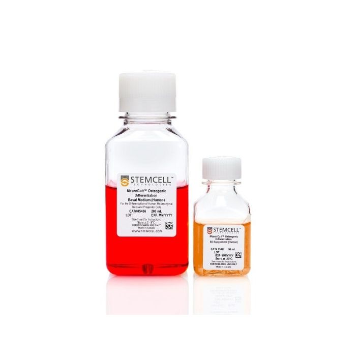
MesenCult™成骨分化基础培养基(人),200 mL MesenCult™成骨分化5X补充剂(人),50 mL

总览

使用MesenCult™成骨分化试剂盒(人)对人间充质基质细胞(MSCs)和hPSC衍生的间充质祖细胞进行高效的成骨分化。该试剂盒可促进在MesenCult™培养基中扩增的MSCs发生显著的矿化和成骨细胞标志物表达,从而能够在各种MSC工作流程中可靠地评估成骨潜能。

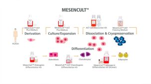
将此试剂盒作为完整的MSC功能评估工作流程的一部分使用。与MesenCult™脂肪生成分化试剂盒(人)以及MesenCult™-ACF软骨生成分化试剂盒配合使用,以评估三谱系分化潜能并验证MSC。这些即用型试剂能够可靠地评估MSC的身份和功能,同时确保在整个MesenCult™工作流程中获得可重复的谱系特异性结果。


细胞类型
间充质干/祖细胞,成骨细胞
 
应用
细胞培养,分化
 
品牌
MesenCult
 
研究领域
药物发现和毒性检测,干细胞生物学
 

实验数据

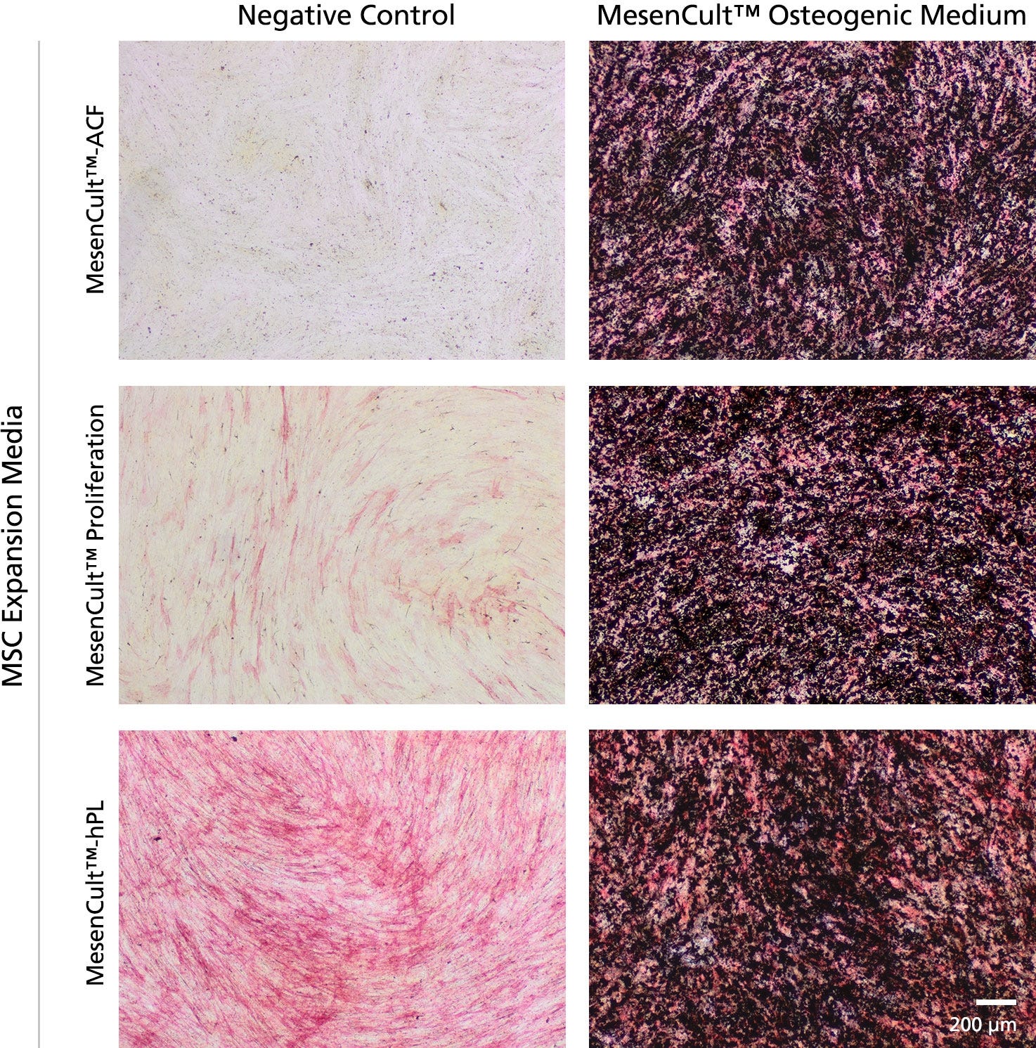
Figure 1. Robust Bone Marrow Mesenchymal Stem and Progenitor Cells (BM MSCs) Osteogenic Differentiation is Achieved in 14 days

Human BM MSCs were derived and expanded for 3 passages using the MesenCult™-ACF Culture Kit (Catalog #05449), MesenCult™ Proliferation Kit (Catalog #05411) or in MesenCult™-hPL Medium (05439). Once MSCs reach greater than 95% confluency, MesenCult™ Osteogenic medium was added to each MSC culture. Osteogenic differentiation was observed within 14 days of induction as indicated by strong alkaline phosphatase activity (red stain) and bone mineralization by the von Kossa method (black stain). Negative controls of undifferentiated MSC cultures were kept in each MesenCult™ MSC expansion media for the same time period. Negative Controls show little or no alkaline phosphatase activity and bone mineralization.

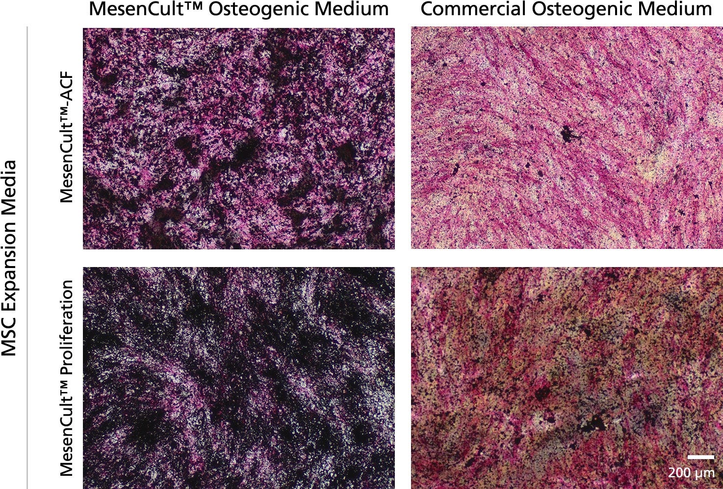
Figure 2. MesenCult™ Osteogenic Medium Leads to Faster and Stronger Osteogenic Differentiation When Compared to A Commercial Osteogenic Differentiation Medium

BM MSCs derived and expanded in MesenCult™-ACF or MesenCult™ Proliferation medium were differentiated for 14 days in either MesenCult™ Osteogenic or another Commercial Osteogenic Medium. Differentiation assays using the MesenCult™ Osteogenic medium displayed stronger alkaline phosphatase activity (red stain) and bone mineralization (black stain) when compared to cultures differentiated with a Commercial Osteogenic Medium.

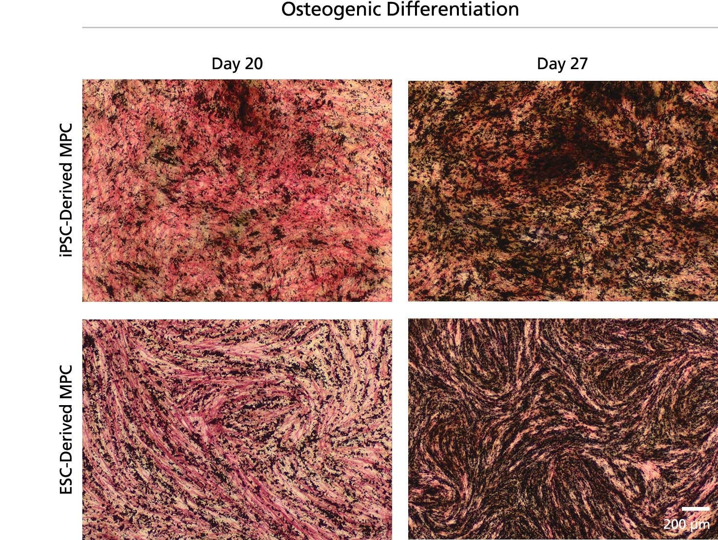
Figure 3. Osteogenic Differentiation of ES-Derived Mesenchymal Progenitor Cells (MPCs)

Mesenchymal progenitor cells (MPCs) were derived from a human iPS or ES cell line using the STEMdiff™ Mesenchymal Progenitor Kit (Catalog #05240) and expanded for 18 or 17 passages, respectively. Cultures of iPS- and ES-derived MPCs were then differentiated for 20 or 27 days in MesenCult™ Osteogenic Differentiation Medium. Strong alkaline phosphatase activity (red stain) and bone mineralization (black stain) were observed at 20 days of osteogenic differentiation, which was further enhanced after 27 days of osteogenic differentiation.

产品说明书及文档

请在《产品说明书》中查找相关支持信息和使用说明,或浏览下方更多实验方案。

Document Type
Product Name
Catalog #
Lot #
Language
Catalog #
05465
Lot #
All
Language
English
Document Type
Safety Data Sheet 1
Catalog #
05465
Lot #
All
Language
English
Document Type
Safety Data Sheet 2
Catalog #
05465
Lot #
All
Language
English

应用领域

本产品专为以下研究领域设计,适用于工作流程中的高亮阶段。探索这些工作流程,了解更多我们为各研究领域提供的其他配套产品。

相关材料与文献

技术资料 (6)

文献 (10)

The Ion Transporter NKCC1 Links Cell Volume to Cell Mass Regulation by Suppressing mTORC1. W. L. Demian et al. Cell reports 2019

Abstract

mTORC1 regulates cellular growth and is activated by growth factors and by essential amino acids such as Leu. Leu enters cells via the Leu transporter LAT1-4F2hc (LAT1). Here we show that the Na+/K+/2Cl- cotransporter NKCC1 (SLC12A2),a known regulator of cell volume,is present in complex with LAT1. We further show that NKCC1 depletion or deletion enhances LAT1 activity,as well as activation of Akt and Erk,leading to activation of mTORC1 in cells,colonic organoids,and mouse colon. Moreover,NKCC1 depletion reduces intracellular Na+ concentration and cell volume (size) and mass and stimulates cell proliferation. NKCC1,therefore,suppresses mTORC1 by inhibiting its key activating signaling pathways. Importantly,by linking ion transport and cell volume regulation to mTORC1 function,NKCC1 provides a long-sought link connecting cell volume (size) to cell mass regulation.
Therapeutic Effects of Mesenchymal Stem Cells Derived From Bone Marrow, Umbilical Cord Blood, and Pluripotent Stem Cells in a Mouse Model of Chemically Induced Inflammatory Bowel Disease. A. Kagia et al. Inflammation 2019 jun

Abstract

Acute inflammatory bowel disease (AIBD) is a wide clinical entity including severe gastrointestinal pathologies with common histopathological basis. Epidemiologically increasing diseases,such as necrotizing enterocolitis (NEC),gastrointestinal graft versus host disease (GVHD),and the primary acute phase of chronic inflammatory bowel disease (CIBD),exhibit a high necessity for new therapeutic strategies. Mesenchymal stem cell (MSC) cellular therapy represents a promising option for the treatment of these diseases. In our study,we comparatively assess the efficacy of human MSCs derived from bone marrow (BM),umbilical cord blood (UCB),human embryonic stem cells (ESCs),or human-induced pluripotent stem cells (iPSCs) in a mouse model of chemically induced acute enterocolitis. The laboratory animals were provided ad libitum potable dextrane sulfate sodium solution (DSS) in order to reproduce an AIBD model and then individually exposed intraperitoneally to MSCs derived from BM (BM-MSCs),UCB (UCB-MSCs),ESCs (ESC-MSCs),or iPSCs (iPSC-MSCs). The parameters used to evaluate the cellular treatment efficacy were the animal survival prolongation and the histopathological-macroscopic picture of bowel sections. Although all categories of mesenchymal stem cells led to statistically significant survival prolongation compared to the control group,significant clinical and histopathological improvement was observed only in mice receiving BM-MSCs and UCB-MSCs. Our results demonstrated that the in vivo anti-inflammatory effect of ESC-MSCs and iPSC-MSCs was inferior to that of UCB-MSCs and BM-MSCs. Further investigation will clarify the potential of ESCs and iPSC-derived MSCs in AIBD treatment.
Mesenchymal stromal cells lower platelet activation and assist in platelet formation in vitro. A. Mendelson et al. JCI insight 2019 aug

Abstract

The complex process of platelet formation originates with the hematopoietic stem cell,which differentiates through the myeloid lineage,matures,and releases proplatelets into the BM sinusoids. How formed platelets maintain a low basal activation state in the circulation remains unknown. We identify Lepr+ stromal cells lining the BM sinusoids as important contributors to sustaining low platelet activation. Ablation of murine Lepr+ cells led to a decreased number of platelets in the circulation with an increased activation state. We developed a potentially novel culture system for supporting platelet formation in vitro using a unique population of CD51+PDGFRalpha+ perivascular cells,derived from human umbilical cord tissue,which display numerous mesenchymal stem cell (MSC) properties. Megakaryocytes cocultured with MSCs had altered LAT and Rap1b gene expression,yielding platelets that are functional with low basal activation levels,a critical consideration for developing a transfusion product. Identification of a regulatory cell that maintains low baseline platelet activation during thrombopoiesis opens up new avenues for improving blood product production ex vivo.
质量保证:

产品仅供研究使用,不用于针对人或动物的诊断或治疗。 欲获悉更多关于STEMCELL的质控信息,请访问 STEMCELL.CN/COMPLIANCE.
Copyright © 2026 by STEMCELL Technologies. All rights reserved.

在线联系