若您需要咨询产品或有任何技术问题,请通过官方电话 400 885 9050 或邮箱 info.cn@stemcell.com 与我们联系。

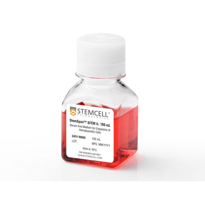
StemSpan™ SFEM II

用于培养和扩增造血细胞的无血清培养基
只有 %1
¥2,380.00

产品号 #(选择产品)

产品号 #09605_C

用于培养和扩增造血细胞的无血清培养基

总览

StemSpan™ 无血清扩增培养基 II(SFEM II)是StemSpan™ SFEM的优化版本,专为体外培养和扩增人造血细胞而开发。该培养基在Iscove’s MDM培养基的基础上包含经过测试的牛血清白蛋白、胰岛素、转铁蛋白以及其他添加物。StemSpan™ SFEM II 中未添加造血细胞生长和扩增所需的重组造血生长因子,这使得用户可以根据实验需求灵活地配制培养基。

搭配合适的细胞因子(例如 StemSpan™ CC100、StemSpan™ CC110 或 StemSpan™ CD34+ 扩增添加物),StemSpan™ SFEM II 可用于扩增来自脐带血、骨髓或其他细胞来源的总有核细胞和 CD34+ 细胞。当与 StemSpan™ 红系扩增添加物(产品号 #02692)、StemSpan™ 髓系扩增添加物(产品号 #02693)、StemSpan™ 髓系扩增添加物 II(产品号 #02694)或 StemSpan™ 巨核细胞扩增添加物(产品号 #02696)分别搭配使用时,StemSpan™ SFEM II 可用于扩增和分化谱系定向祖细胞,以生成有核红细胞、粒细胞、单核细胞或巨核细胞。

包含
• Iscove’s MDM
• 牛血清白蛋白
• 重组人胰岛素
• 人转铁蛋白(铁饱和)
• 2-巯基乙醇
• 添加物
 
分类
专用培养基
 
细胞类型
造血干/祖细胞
 
种属

 
应用
细胞培养,扩增
 
品牌
StemSpan
 
研究领域
药物发现和毒性检测,干细胞生物学,移植研究
 
制剂类别
无血清
 

实验数据

Expansion of CD34 + Human Cord Blood Cells Cultured in StemSpan™ Media Containing CC100 Cytokine Cocktail

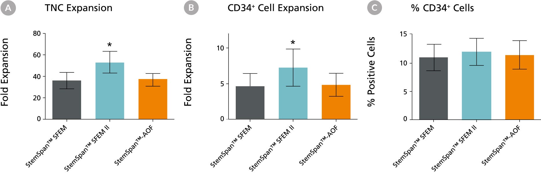
Figure 1. Expansion of CD34+ Human Cord Blood Cells Cultured in StemSpan™ Media Containing CC100 Cytokine Cocktail

Purified CD34+ human cord blood (CB) cells were suspended at a concentration of 10,000 per mL in StemSpan™ SFEM (dark gray bars), SFEM II (blue bars) and AOF (orange bars) media containing CC100 Cytokine Cocktail (Catalog #02690). Cultures were maintained for 7 days, after which the cells were counted and examined for CD34 and CD45 expression by flow cytometry. Shown are the fold expansion of total nucleated cells (TNC) (A) and CD34+ cells (B) per input CD34+ cell, and the percent CD34 + cells (C). Results represent the average results of 32 different CB samples. Vertical lines indicate 95% confidence limits, the range within which 95% of results fall. The numbers of cells produced in StemSpan™ SFEM II were significantly higher than in StemSpan™ SFEM and StemSpan™-AOF (*p<0.001, paired t-test, n=32).

Note: Data for StemSpan™-AOF shown were generated with the original phenol red-containing version StemSpan™-ACF (Catalog #09855). However internal testing showed that the performance of the new phenol red-free, cGMP-manufactured version, StemSpan™-AOF (Catalog #100-0130) was comparable.

StemSpan™ SFEM II Serum-Free Expansion Medium Containing CC100 Cytokine Cocktail Supports Greater Expansion of Human CD34 + Cells Than Other Media Tested

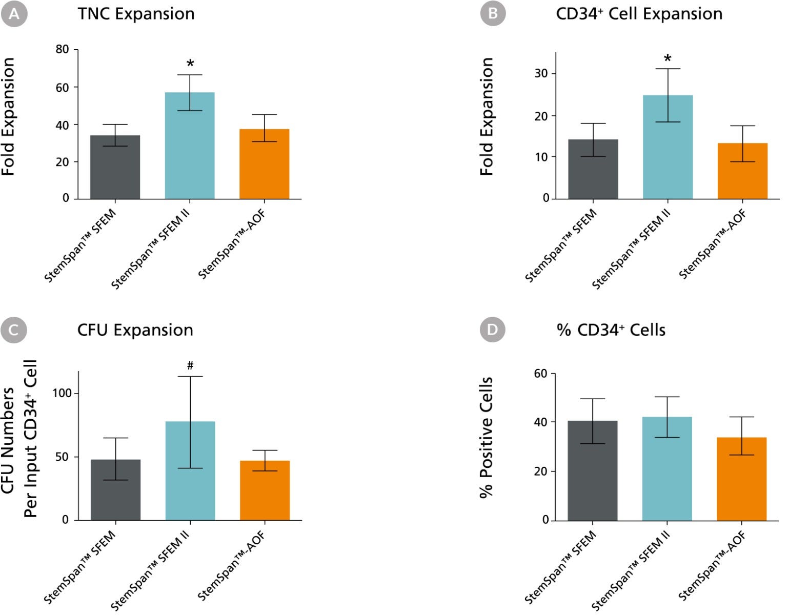
Figure 2. Expansion of CD34+ Human Cord Blood Cells Cultured in StemSpan™ Media Containing CD34+ Expansion Supplement

Purified CD34+ human cord blood (CB) cells were suspended at a concentration of 10,000 per mL in StemSpan™ SFEM (dark gray bars), SFEM II (blue bars) and AOF (orange bars) media containing CD34+ Expansion Supplement (Catalog #02691). Cultures were maintained for 7 days, after which the cells were counted and examined for CD34 and CD45 expression by flow cytometry. The number of colony-forming units (CFU) in the expanded population was determined by replating cells in MethoCult™ H4435 and counting the number of colonies produced 14 days later. Shown are the fold expansion of total nucleated cells (TNC) (A), CD34+ cells (B) and CFU numbers (C) per input CD34+ cell, and the percent CD34+ cells (D) in these cultures (n=6). Vertical lines indicate 95% confidence limits, the range within which 95% of results fall. The numbers of cells produced in StemSpan™ SFEM II was significantly higher than in SFEM and AOF (*p<0.001, #p<0.05, paired t-test, n=6).

Note: Data for StemSpan™-AOF shown were generated with the original phenol red-containing version StemSpan™-ACF (Catalog #09855). However internal testing showed that the performance of the new phenol red-free, cGMP-manufactured version, StemSpan™-AOF (Catalog #100-0130) was comparable.

Expansion of CD34 + Human Cord Blood Cells Cultured in StemSpan™ Media Containing CD34 + Expansion Supplement

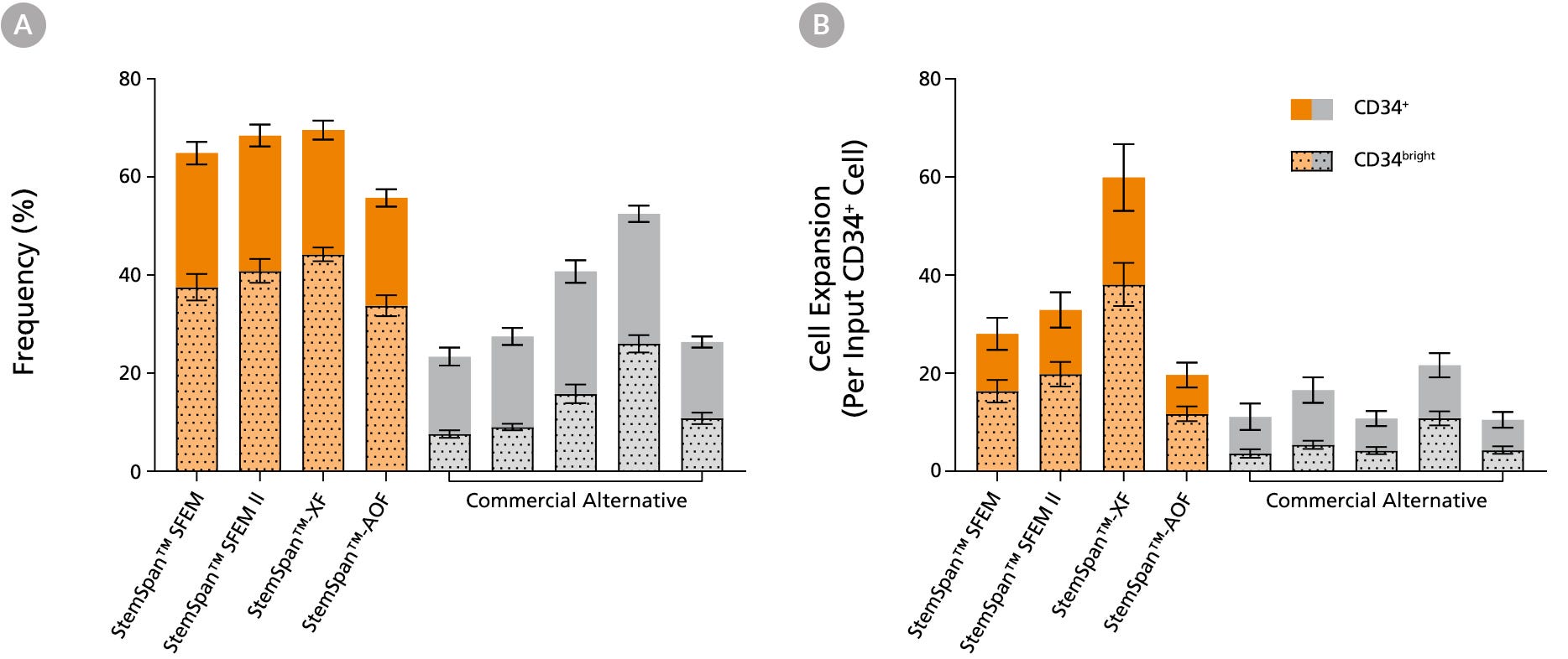
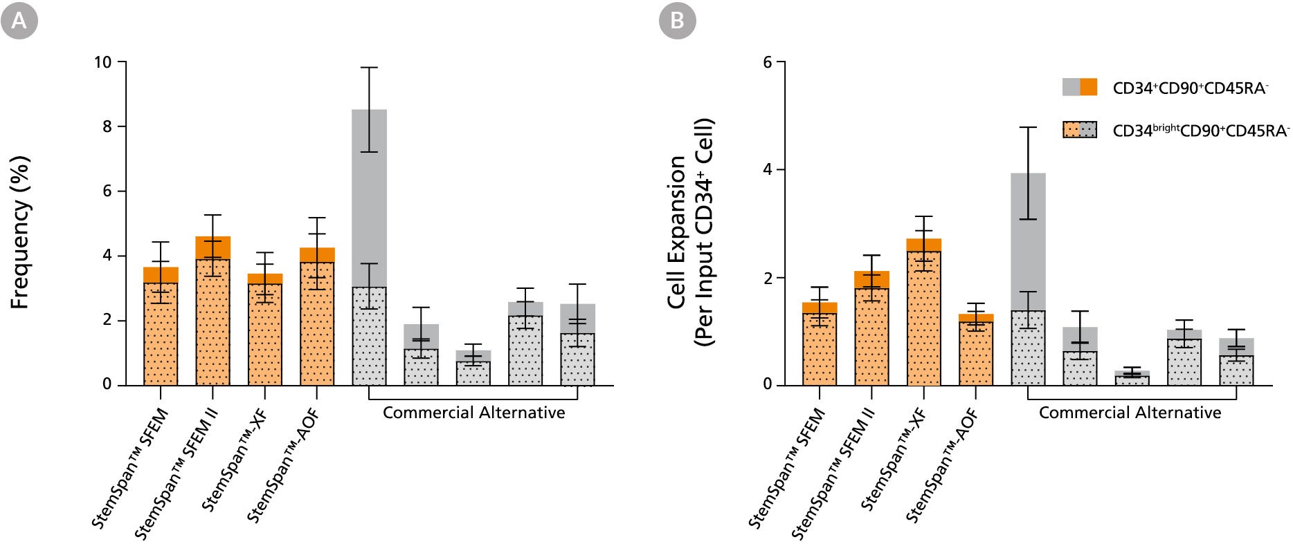
Figure 3. StemSpan™ Media Support Greater Expansion of Human CD34+ and CD34bright Cells than Other Commercial Media

Purified CB-derived CD34+ cells were cultured for 7 days in select StemSpan™ media (StemSpan™ SFEM, StemSpan™ SFEM II, StemSpan™-XF, or StemSpan™-AOF, orange bars), and in five xeno-free media formulations from other suppliers (Xeno-Free Commercial Alternative, grey bars) including (in random order) CTS™ StemPro™ HSC (Thermo), SCGM (Cellgenix), X-VIVO™ 15 (Lonza), Stemline™ II (Sigma), and StemPro™-34 (Thermo). All media were supplemented with StemSpan™ CD34+ Expansion Supplement and UM171*. The (A) frequency and (B) cell expansion of viable CD34+ and CD34bright cells in culture were based on viable cell counts and flow cytometry results as shown in Figure 1. StemSpan™ showed significantly higher expansion of CD34+ and CD34bright cells (P < 0.05 when comparing StemSpan™ SFEM II to five media from other suppliers, calculated using a one-way ANOVA followed by Dunnett’s post hoc test) and StemSpan™-AOF, the only animal origin-free formulation, showed equivalent performance to all xeno-free commericals alternatives tested. Data shown are mean ± SEM (n = 8).

Note: Data for StemSpan™-AOF shown were generated with the original phenol red-containing version StemSpan™-ACF (Catalog #09855). However internal testing showed that the performance of the new phenol red-free, cGMP-manufactured version, StemSpan™-AOF (Catalog #100-0130) was comparable. *Similar results are expected when using UM729 (Catalog #72332) prepared to a final concentration of 1μM. For more information including data comparing UM171 and UM729, see Fares et al., 2014.

StemSpan™ SFEM II Serum-Free Expansion Medium Containing Megakaryocyte Expansion Supplement Supports Greater Expansion of Megakaryocytes Than Other Media Tested

Figure 4. StemSpan™ Media Support Equal or Greater Expansion of Primitive Human CD34brightCD90+CD45RA- Cells Than Other Commercial Media

Purified CB-derived CD34+ cells were cultured for 7 days in select StemSpan™ media (StemSpan™ SFEM, StemSpan™ SFEM II, StemSpan™-XF, or StemSpan™-AOF, orange bars), and in five xeno-free media formulations from other suppliers (Commercial Alternative, grey bars) including (in random order) CTS StemPro HSC (Thermo), SCGM (Cellgenix), X-VIVO 15 (Lonza), Stemline II (Sigma), and StemPro 34 (Thermo). All media were supplemented with StemSpan™ CD34+ Expansion Supplement and UM171*. The (A) frequency and (B) cell expansion of CD34+CD90+CD45RA- (solid) and CD34brightCD90+CD45RA-(dotted overlay) cells in culture were based on viable cell counts and flow cytometry results as shown in Figure 1. StemSpan™ media showed similar or significantly higher expansion of CD34brightCD90+CD45RA- cells (P < 0.05 compared to five media from other suppliers, calculated using one-way ANOVA followed by Dunnett’s post hoc test) and StemSpan™-AOF, the only animal origin-free formulation tested, showed equivalent performance to all xeno-free commercial alternatives tested. Data shown are mean ± SEM (n = 8).

Note: Data for StemSpan™-AOF shown were generated with the original phenol red-containing version StemSpan™-ACF (Catalog #09855). However internal testing showed that the performance of the new phenol red-free, cGMP-manufactured version, StemSpan™-AOF (Catalog #100-0130) was comparable.

*Similar results are expected when using UM729 (Catalog #72332) prepared to a final concentration of 1μM. For more information including data comparing UM171 and UM729, see Fares et al. 2014.

StemSpan™ SFEM II Serum-Free Expansion Medium Containing Megakaryocyte Expansion Supplement Supports Greater Expansion of Megakaryocytes Than Other Media Tested

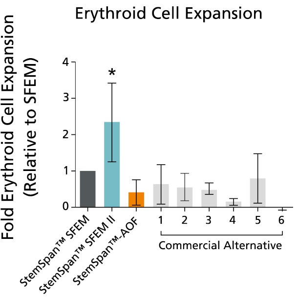
Figure 5. StemSpan™ SFEM II Serum-Free Expansion Medium Containing Erythroid Expansion Supplement Supports Greater Expansion of Erythroid Cells Than Other Media Tested

The numbers of erythroid cells, normalized relative to the values obtained in StemSpan™ SFEM medium (dark gray bar), obtained after culturing purified CD34+ CB cells for 14 days in StemSpan™ SFEM, SFEM II (blue bar) and AOF (orange bar), and six media from other commercial suppliers (light gray bars, commercial alternative 1-6, which included, in random order, X-Vivo-15 and HPGM (both from Lonza), StemLine II (Sigma), HP01 (Macopharma), StemPro34 (Life Technologies) and SCGM (Cellgenix). All media were supplemented with StemSpan™ Erythroid Expansion Supplement (Catalog #02692). Vertical lines indicate 95% confidence limits, the range within which 95% of results fall. The numbers of cells produced in StemSpan™ SFEM II were significantly higher than in all other media (*p<0.05, paired t-test, n=6).

Note: Data for StemSpan™-AOF shown were generated with the original phenol red-containing version StemSpan™-ACF (Catalog #09855). However internal testing showed that the performance of the new phenol red-free, cGMP-manufactured version, StemSpan™-AOF (Catalog #100-0130) was comparable.

StemSpan™ SFEM II Serum-Free Expansion Medium Containing Megakaryocyte Expansion Supplement Supports Greater Expansion of Megakaryocytes Than Other Media Tested

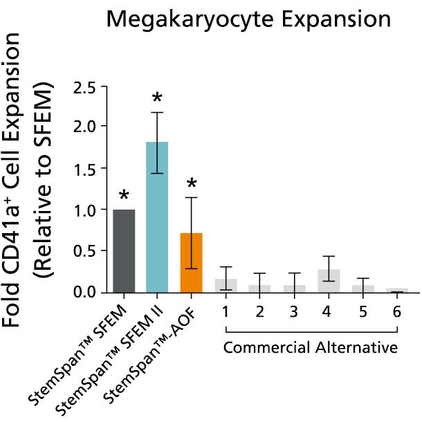
Figure 6. StemSpan™ SFEM II Serum-Free Expansion Medium Containing Megakaryocyte Expansion Supplement Supports Greater Expansion of Megakaryocytes Than Other Media Tested

The numbers of megakaryocytes, normalized relative to the values obtained in StemSpan™ SFEM medium (dark gray bar), obtained after culturing purified CD34+ CB cells for 14 days in StemSpan™ SFEM, SFEM II (blue bar) and AOF (orange bar), and six media from other commercial suppliers (light gray bars, Commercial Alternative 1-6, which included, in random order, StemLine II (Sigma), HPGM (Lonza), HP01 (Macopharma), SCGM (Cellgenix), StemPro34 (Life Technologies) and X-Vivo-15 (Lonza). All media were supplemented with StemSpan™ Megakaryocyte Expansion Supplement (Catalog #02696). Vertical lines indicate 95% confidence limits, the range within which 95% of results fall. The numbers of cells produced in the StemSpan™ media were significantly higher than in the other media (*p<0.01 paired t-test, n=6).

Note: Data for StemSpan™-AOF shown were generated with the original phenol red-containing version StemSpan™-ACF (Catalog #09855). However internal testing showed that the performance of the new phenol red-free, cGMP-manufactured version, StemSpan™-AOF (Catalog #100-0130) was comparable.

Table 1. Production of Myeloid Cells from Human CB CD34+ Cells Cultured in SFEM II Containing Myeloid Expansion Supplement or Myeloid Expansion Supplement ll

StemSpan™ SFEM II Serum-Free Expansion Medium Containing Erythroid Expansion Supplement Supports Greater Expansion of Erythroid Cells Than Other Media Tested

Shown are numbers of total nucleated cells (TNCs) produced per input human CB-derived CD34+ cell and percentages of cells positive for myeloid markers CD13, CD14 and CD15 after 14 days of culture in SFEM II containing Myeloid Expansion Supplement (n = 14) or Myeloid Expansion Supplement II (n = 16). *95% confidence limits (CL); the range within which 95% of results typically fall.

产品说明书及文档

请在《产品说明书》中查找相关支持信息和使用说明,或浏览下方更多实验方案。

Document Type
Product Name
Catalog #
Lot #
Language
Catalog #
09605, 09655
Lot #
All
Language
English
Document Type
Safety Data Sheet
Catalog #
09605, 09655
Lot #
All
Language
English

应用领域

本产品专为以下研究领域设计,适用于工作流程中的高亮阶段。探索这些工作流程,了解更多我们为各研究领域提供的其他配套产品。

相关材料与文献

技术资料 (27)

文献 (60)

Technical advance: ascorbic acid induces development of double-positive T cells from human hematopoietic stem cells in the absence of stromal cells. Huijskens MJAJ et al. Journal of leukocyte biology 2014 DEC

Abstract

The efficacy of donor HSCT is partly reduced as a result of slow post-transplantation immune recovery. In particular,T cell regeneration is generally delayed,resulting in high infection-related mortality in the first years post-transplantation. Adoptive transfer of in vitro-generated human T cell progenitors seems a promising approach to accelerate T cell recovery in immunocompromised patients. AA may enhance T cell proliferation and differentiation in a controlled,feeder-free environment containing Notch ligands and defined growth factors. Our experiments show a pivotal role for AA during human in vitro T cell development. The blocking of NOS diminished this effect,indicating a role for the citrulline/NO cycle. AA promotes the transition of proT1 to proT2 cells and of preT to DP T cells. Furthermore,the addition of AA to feeder cocultures resulted in development of DP and SP T cells,whereas without AA,a preT cell-stage arrest occurred. We conclude that neither DLL4-expressing feeder cells nor feeder cell conditioned media are required for generating DP T cells from CB and G-CSF-mobilized HSCs and that generation and proliferation of proT and DP T cells are greatly improved by AA. This technology could potentially be used to generate T cell progenitors for adoptive therapy.
Selection-free genome editing of the sickle mutation in human adult hematopoietic stem/progenitor cells. M. A. DeWitt et al. Science translational medicine 2016 OCT

Abstract

Genetic diseases of blood cells are prime candidates for treatment through ex vivo gene editing of CD34+ hematopoietic stem/progenitor cells (HSPCs),and a variety of technologies have been proposed to treat these disorders. Sickle cell disease (SCD) is a recessive genetic disorder caused by a single-nucleotide polymorphism in the $\beta$-globin gene (HBB). Sickle hemoglobin damages erythrocytes,causing vasoocclusion,severe pain,progressive organ damage,and premature death. We optimize design and delivery parameters of a ribonucleoprotein (RNP) complex comprising Cas9 protein and unmodified single guide RNA,together with a single-stranded DNA oligonucleotide donor (ssODN),to enable efficient replacement of the SCD mutation in human HSPCs. Corrected HSPCs from SCD patients produced less sickle hemoglobin RNA and protein and correspondingly increased wild-type hemoglobin when differentiated into erythroblasts. When engrafted into immunocompromised mice,ex vivo treated human HSPCs maintain SCD gene edits throughout 16 weeks at a level likely to have clinical benefit. These results demonstrate that an accessible approach combining Cas9 RNP with an ssODN can mediate efficient HSPC genome editing,enables investigator-led exploration of gene editing reagents in primary hematopoietic stem cells,and suggests a path toward the development of new gene editing treatments for SCD and other hematopoietic diseases.
CRISPR/Cas9 $\beta$-globin gene targeting in human haematopoietic stem cells. D. P. Dever et al. Nature 2016 NOV

Abstract

The $\beta$-haemoglobinopathies,such as sickle cell disease and $\beta$-thalassaemia,are caused by mutations in the $\beta$-globin (HBB) gene and affect millions of people worldwide. Ex vivo gene correction in patient-derived haematopoietic stem cells followed by autologous transplantation could be used to cure $\beta$-haemoglobinopathies. Here we present a CRISPR/Cas9 gene-editing system that combines Cas9 ribonucleoproteins and adeno-associated viral vector delivery of a homologous donor to achieve homologous recombination at the HBB gene in haematopoietic stem cells. Notably,we devise an enrichment model to purify a population of haematopoietic stem and progenitor cells with more than 90{\%} targeted integration. We also show efficient correction of the Glu6Val mutation responsible for sickle cell disease by using patient-derived stem and progenitor cells that,after differentiation into erythrocytes,express adult $\beta$-globin (HbA) messenger RNA,which confirms intact transcriptional regulation of edited HBB alleles. Collectively,these preclinical studies outline a CRISPR-based methodology for targeting haematopoietic stem cells by homologous recombination at the HBB locus to advance the development of next-generation therapies for $\beta$-haemoglobinopathies.

更多信息

更多信息
物种
Contains • Iscove’s MDM • Bovine serum albumin • Recombinant human insulin • Human transferrin (iron-saturated) • 2-Mercaptoethanol • Supplements
配方 无血清
质量保证:

产品仅供研究使用,不用于针对人或动物的诊断或治疗。 欲获悉更多关于STEMCELL的质控信息,请访问 STEMCELL.CN/COMPLIANCE.
Copyright © 2026 by STEMCELL Technologies. All rights reserved.

在线联系