若您需要咨询产品或有任何技术问题,请通过官方电话 400 885 9050 或邮箱 info.cn@stemcell.com 与我们联系。

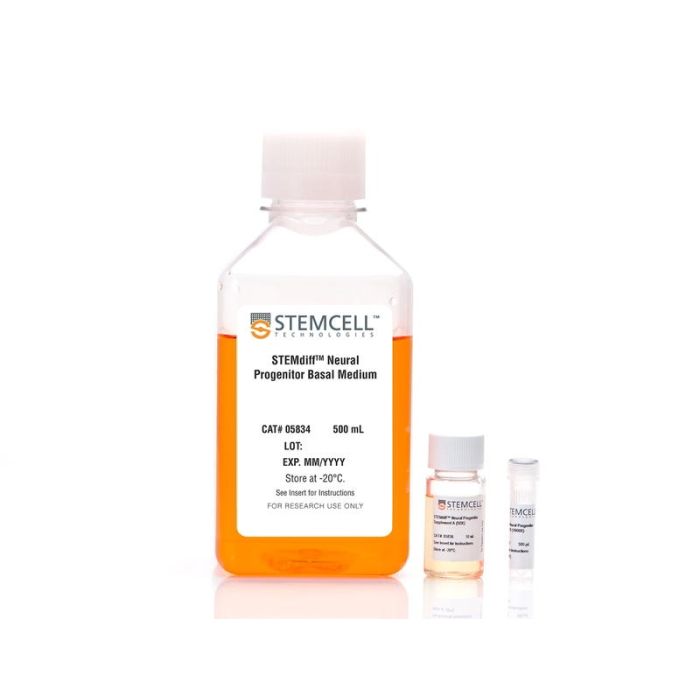
STEMdiff™神经前体细胞培养基

用于维持和扩增人ES和iPS细胞衍生的神经前体细胞的培养基
只有 %1
¥4,562.00

产品号 #(选择产品)

产品号 #05833_C

用于维持和扩增人ES和iPS细胞衍生的神经前体细胞的培养基

产品优势

  • 成分明确且不含血清
  • 支持使用 STEMdiff™ 神经诱导培养基生成的NPC的扩增
  • 针对多代传代过程中的神经祖细胞高效扩增进行了优化
  • 保留NPC的多能性,同时最大程度地减少自发神经元分化
  • 使用方便,配方和操作流程简明

产品组分包括

  • STEMdiff™ 神经前体细胞基础培养基,500 mL
  • STEMdiff™ 神经前体细胞补充剂 A (50X),10 mL
  • STEMdiff™ 神经前体细胞补充剂 B (1000X),500 µL

总览

STEMdiff™ 神经前体细胞培养基是一种成分明确且不含血清的培养基,适用于扩增使用 STEMdiff™ 神经诱导培养基(目录号 #05835)从人胚胎干细胞(ES)和诱导多能干细胞(iPS)诱导获得的神经前体细胞(NPCs)。在该培养基中培养的NPCs每代可扩增3–5倍,且可连续传代至少10代,同时自发性神经元分化极少。

分类
专用培养基
 
细胞类型
神经细胞,PSC衍生,神经干/祖细胞,多能干细胞
 
种属

 
应用
细胞培养,扩增
 
品牌
STEMdiff
 
研究领域
疾病建模,药物发现和毒理检测,神经科学,干细胞生物学
 
制剂类别
无血清
 

实验数据

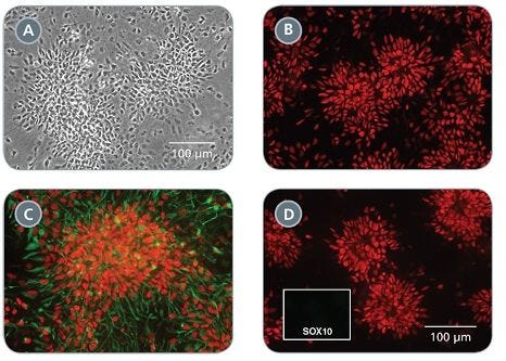
Morphology and Marker Expression of Neural Progenitor Cells Cultured in STEMdiff™ Neural Progenitor Medium

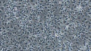
Figure 1. Morphology and Marker Expression of Neural Progenitor Cells Cultured in STEMdiff™ Neural Progenitor Medium

(A) Typical NPC morphology is observed in cultures (shown at day 6 of passage 1). (B-D) NPCs maintained in STEMdiff™ Neural Progenitor Medium express the CNS-type NPC markers PAX6 (B, D, red), SOX1 (C, red) and NESTIN (C, green), but not the neural crest marker SOX10 (D, green, single channel shown in inset). B-D were taken at the same magnification.

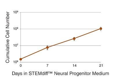
Expansion of Neural Progenitor Cells in STEMdiff™ Neural Progenitor Medium

Figure 2. Expansion of Neural Progenitor Cells in STEMdiff™ Neural Progenitor Medium

NPCs cultured in STEMdiff™ Neural Progenitor Medium can be expanded to generate a large number of cells. Three- to five-fold expansion can be achieved upon each passage. NPCs were derived using STEMdiff™ Neural Induction Medium and passaged once a week on average. n = 6.

Neural Progenitor Cells Cultured in STEMdiff™ Neural Progenitor Medium Show Minimal Spontaneous Neuronal Differentiation

Figure 3. Neural Progenitor Cells Cultured in STEMdiff™ Neural Progenitor Medium Show Minimal Spontaneous Neuronal Differentiation

Passages 1 (A) and 3 (B) of a representative NPC culture maintained in STEMdiff™ Neural Progenitor Medium. Cells were immunolabeled with SOX1 (red) to identify NPCs, and class III β-tubulin (green) to identify neurons. Spontaneous neuronal differentiation is low in NPC cultures maintained in STEMdiff™ Neural Progenitor Medium. A and B were taken at the same magnification.

Neural Progenitor Cells Maintained in STEMdiff™ Neural Progenitor Medium can Differentiate into Neurons and Astrocytes

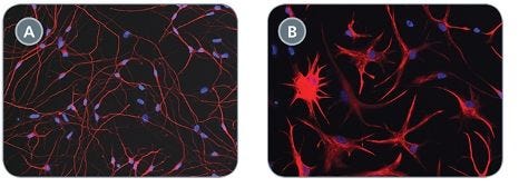
Figure 4. Neural Progenitor Cells Maintained in STEMdiff™ Neural Progenitor Medium can Differentiate into Neurons and Astrocytes

When directed according to published protocols, NPCs can differentiate into neurons (A, class III β-tubulin shown in red) and astrocytes (B, GFAP shown in red). Nuclei are counterstained with DAPI (blue).

产品说明书及文档

请在《产品说明书》中查找相关支持信息和使用说明,或浏览下方更多实验方案。

Document Type
Product Name
Catalog #
Lot #
Language
Catalog #
05833
Lot #
All
Language
English
Document Type
Technical Manual
Catalog #
05833
Lot #
All
Language
English
Document Type
Safety Data Sheet 1
Catalog #
05833
Lot #
All
Language
English
Document Type
Safety Data Sheet 2
Catalog #
05833
Lot #
All
Language
English
Document Type
Safety Data Sheet 3
Catalog #
05833
Lot #
All
Language
English

应用领域

本产品专为以下研究领域设计,适用于工作流程中的高亮阶段。探索这些工作流程,了解更多我们为各研究领域提供的其他配套产品。

相关材料与文献

技术资料 (18)

文献 (31)

Multisystemic Disease Modeling of Liver-Derived Protein Folding Disorders Using Induced Pluripotent Stem Cells (iPSCs). Leung A and Murphy GJ Methods in molecular biology (Clifton,N.J.) 2016 JAN

Abstract

Familial transthyretin amyloidosis (ATTR) is an autosomal dominant protein-folding disorder caused by over 100 distinct mutations in the transthyretin (TTR) gene. In ATTR,protein secreted from the liver aggregates and forms fibrils in target organs,chiefly the heart and peripheral nervous system,highlighting the need for a model capable of recapitulating the multisystem complexity of this clinically variable disease. Here,we describe detailed methodologies for the directed differentiation of protein folding disease-specific iPSCs into hepatocytes that produce mutant protein,and neural-lineage cells often targeted in disease. Methodologies are also described for the construction of multisystem models and drug screening using iPSCs.
Genome modification leads to phenotype reversal in human myotonic dystrophy type 1 induced pluripotent stem cell-derived neural stem cells. Xia G et al. Stem cells (Dayton,Ohio) 2015 JUN

Abstract

Myotonic dystrophy type 1 (DM1) is caused by expanded CTG repeats in the 3'-untranslated region (3' UTR) of the DMPK gene. Correcting the mutation in DM1 stem cells would be an important step toward autologous stem cell therapy. The objective of this study is to demonstrate in vitro genome editing to prevent production of toxic mutant transcripts and reverse phenotypes in DM1 stem cells. Genome editing was performed in DM1 neural stem cells (NSCs) derived from human DM1 induced pluripotent stem (iPS) cells. An editing cassette containing SV40/bGH polyA signals was integrated upstream of the CTG repeats by TALEN-mediated homologous recombination (HR). The expression of mutant CUG repeats transcript was monitored by nuclear RNA foci,the molecular hallmarks of DM1,using RNA fluorescence in situ hybridization. Alternative splicing of microtubule-associated protein tau (MAPT) and muscleblind-like (MBNL) proteins were analyzed to further monitor the phenotype reversal after genome modification. The cassette was successfully inserted into DMPK intron 9 and this genomic modification led to complete disappearance of nuclear RNA foci. MAPT and MBNL 1,2 aberrant splicing in DM1 NSCs were reversed to normal pattern in genome-modified NSCs. Genome modification by integration of exogenous polyA signals upstream of the DMPK CTG repeat expansion prevents the production of toxic RNA and leads to phenotype reversal in human DM1 iPS-cells derived stem cells. Our data provide proof-of-principle evidence that genome modification may be used to generate genetically modified progenitor cells as a first step toward autologous cell transfer therapy for DM1.
Generation of Functional Cardiomyocytes from Efficiently Generated Human iPSCs and a Novel Method of Measuring Contractility. Rajasingh S et al. PloS one 2015 AUG

Abstract

Human induced pluripotent stem cells (iPSCs) derived cardiomyocytes (iCMCs) would provide an unlimited cell source for regenerative medicine and drug discoveries. The objective of our study is to generate functional cardiomyocytes from human iPSCs and to develop a novel method of measuring contractility of CMCs. In a series of experiments,adult human skin fibroblasts (HSF) and human umbilical vein endothelial cells (HUVECs) were treated with a combination of pluripotent gene DNA and mRNA under specific conditions. The iPSC colonies were identified and differentiated into various cell lineages,including CMCs. The contractile activity of CMCs was measured by a novel method of frame-by-frame cross correlation (particle image velocimetry-PIV) analysis. Our treatment regimen transformed 4% of HSFs into iPSC colonies at passage 0,a significantly improved efficiency compared with use of either DNA or mRNA alone. The iPSCs were capable of differentiating both in vitro and in vivo into endodermal,ectodermal and mesodermal cells,including CMCs with<88% of cells being positive for troponin T (CTT) and Gata4 by flow cytometry. We report a highly efficient combination of DNA and mRNA to generate iPSCs and functional iCMCs from adult human cells. We also report a novel approach to measure contractility of iCMCs.

更多信息

更多信息
物种
配方 无血清
质量保证:

产品仅供研究使用,不用于针对人或动物的诊断或治疗。 欲获悉更多关于STEMCELL的质控信息,请访问 STEMCELL.CN/COMPLIANCE.
Copyright © 2026 by STEMCELL Technologies. All rights reserved.

在线联系