若您需要咨询产品或有任何技术问题,请通过官方电话 400 885 9050 或邮箱 info.cn@stemcell.com 与我们联系。

STEMdiff™肠道类器官试剂盒

用于从 hPSC 建立和维持人肠道类器官的无血清培养基试剂盒
只有 %1
¥16,304.00

产品号 #(选择产品)

产品号 #05140


用于从 hPSC 建立和维持人肠道类器官的无血清培养基试剂盒

产品优势

  • 适用于发育中小肠上皮及相关间质的相关模型系统
  • 高效培养基支持人胚胎干细胞(ES)和诱导多能干细胞(iPS)高效分化为肠类器官
  • 方便的模型系统,可通过传代长期维持或冷冻保存,提供实验灵活性
  • 无血清培养基体系经过优化,减少实验变异性

产品组分包括

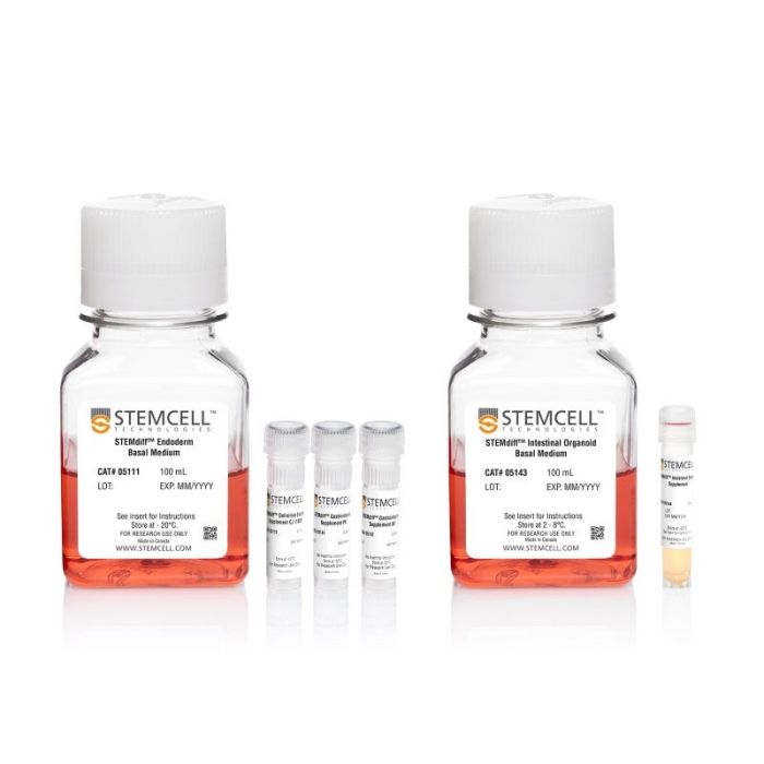
  • STEMdiff™ 内胚层基础培养基,100 mL
  • STEMdiff™ 定型内胚层补充剂 CJ,1.1 mL
  • STEMdiff™ 胃肠补充剂 PK,0.64 mL
  • STEMdiff™ 胃肠补充剂 UB,0.64 mL
  • STEMdiff™ 肠道类器官基础培养基 ,100 mL
  • STEMdiff™ 肠道类器官补充剂,2 mL
Need a high-quality cell source? Use the hiPSC SCTi003-A (female) or SCTi004-A (male) control lines, manufactured with mTeSR™ Plus.
专为您的实验方案打造的产品
要查看实验方案所需的所有配套产品,请参阅《实验方案与技术文档》

总览

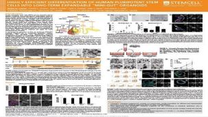
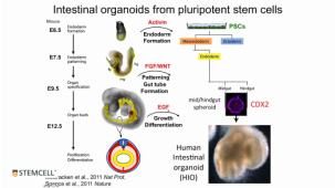
STEMdiff™肠类器官试剂盒(产品号 #05140)是一种无血清的细胞培养体系,可通过简单的三阶段方案高效、稳定地生成源自人多能干细胞(hPSC)的肠类器官。使用该试剂盒,hPSCs可依次诱导生成确定性内胚层、中/后肠球状体及肠类器官,这些类器官可长期传代培养或冻存。生成的肠类器官具有模拟发育中肠上皮及相关间质的细胞组成和组织结构,是一个与肠发育直接相关的便捷模型系统。STEMdiff™肠类器官试剂盒基于Spence等人(Nature 2011)发表的配方,经过优化以提高不同hPSC系间类器官形成和扩增的效率与一致性。人肠类器官可作为研究肠发育、细胞生物学、肠炎、肠再生、微生物相互作用、疾病建模、药物发现及化合物筛选的模型系统。

该试剂盒针对在mTeSR™1、mTeSR™ Plus或TeSR™-AOF中培养的细胞进行了优化。

如需长期培养和传代,类器官培养所需的试剂盒组分可作为 STEMdiff™ 肠道类器官生长培养基(产品号 #05145)单独购买。

细胞类型
内胚层,PSC衍生,肠道细胞
 
种属

 
应用
细胞培养,分化,类器官培养
 
品牌
STEMdiff
 
研究领域
疾病建模,药物发现和毒理检测,上皮细胞研究
 
制剂类别
无血清
 

实验数据

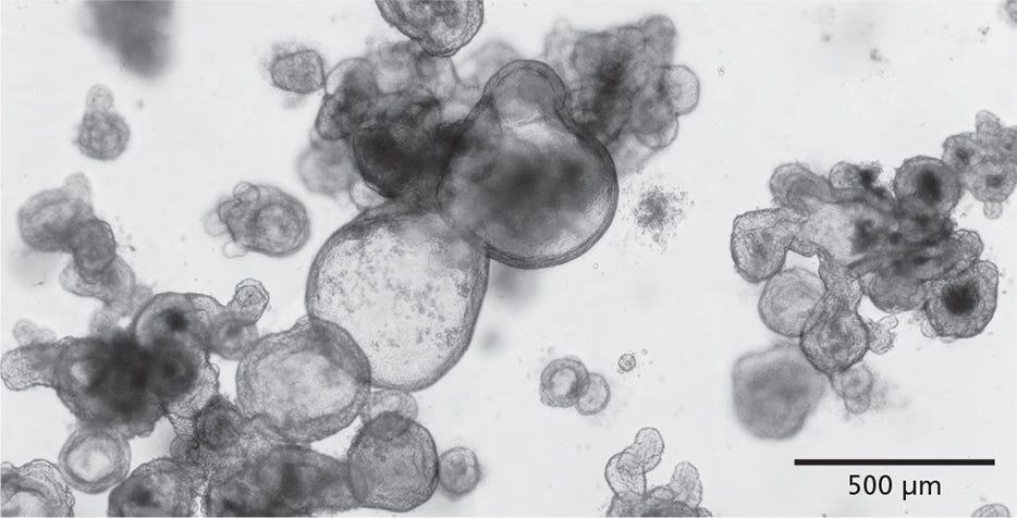
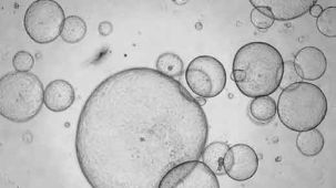
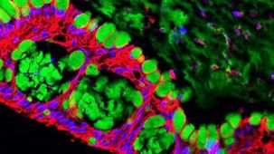
Figure 1. STEMdiff™ Intestinal Organoid Kit Enables the Growth of Intestinal Organoids from PSCs

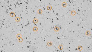
STEMdiff™ Intestinal Organoid Kit facilitates directed differentiation of PSCs to form human small intestinal organoids. The organoids exhibit a polarized epithelial monolayer surrounding a hollow lumen and are associated with a mesenchymal cell population. Pictured are passage 3 human intestinal organoids generated using this kit.

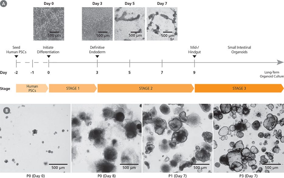
Figure 2. Generation of Human Intestinal Organoid Cultures Using the STEMdiff™ Intestinal Organoid Kit

(A) Human PSC cultures progress through a three-stage differentiation process to generate human intestinal organoids. By day 3 of the protocol, cultures exhibit characteristics typical of definitive endoderm and mid-/hindgut differentiation is initiated. During mid-/hindgut differentiation (days 5 - 9), cells form mid-/hindgut spheroids that are released from the cell monolayer into the culture medium. These spheroids are collected and embedded in extracellular matrix. (B) Embedded mid-/hindgut spheroids cultured in STEMdiff™ Intestinal Organoid Growth Medium mature into intestinal organoids (days in parentheses indicate days post-embedding in a given passage). Once established, intestinal organoids can be maintained and expanded in culture by passaging every 7 - 10 days. After multiple passages, the organoids generally exhibit less sinking within the matrix dome and a lower proportion of mesenchymal cells.

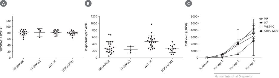
Figure 3. STEMdiff™ Intestinal Organoid Kit Supports Robust Differentiation and Expansion Across ESC and iPSC Lines

STEMdiff™ Intestinal Organoid Kit enables high-efficiency generation of intestinal organoids from both ESCs (H9, H7) and iPSCs (WLS-1C, STiPS-MOO1). (A) Organoids initiated from a variety of cell lines show efficient induction of definitive endoderm, measured by co-expression of FOXA2 and SOX17 on day 3 of differentiation. (B) Both ESCand iPSC-derived cultures demonstrate efficient spheroid formation upon mid-/hindgut induction. The total number of spheroids obtained per well in a given differentiation is shown. (C) Organoids cultured from either ESCs or iPSCs can be expanded and maintained over multiple passages. Shown is the total cell yield per passage. Organoids were passaged every 7 - 10 days with a split ratio between 1 in 2 and 1 in 4. Data points represent the mean of 3 biological replicates. Error bars throughout represent standard deviation of the mean.

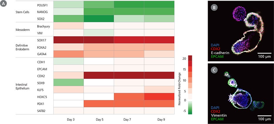
Figure 4. Characteristics of Mid-/Hindgut Spheroids Generated with STEMdiff™ Intestinal Organoid Kit

(A) Cultures differentiated using STEMdiff™ Intestinal Organoid Kit exhibit the expected markers during definitive endoderm and mid-/hindgut specification. During the protocol, gene expression patterns shift from pluripotency markers (day 0) to definitive endoderm markers by day 3 and those of the mid-/hindgut epithelium by day 9. Mid-/hindgut cultures (day 9) also express markers of the associated mesenchyme. Marker levels were assessed by RT-qPCR and normalized to expression levels for undifferentiatied H9 cells. (B) Mid-/hindgut spheroids (day 9) express markers of the intestinal epithelium (CDX2, E-cadherin, EPCAM). (C) Mid-/hindgut spheroids (day 9) also incorporate components of the associated mesenchyme (vimentin).

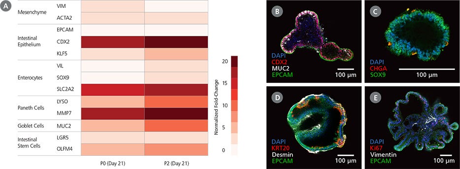
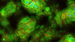
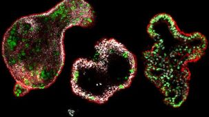
Figure 5. Intestinal Organoids Cultured In STEMdiff™ Intestinal Organoid Growth Medium Exhibit Features of the Intestinal Epithelium

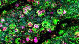
(A) Differentiated PSC-derived intestinal organoids express markers of the intestinal epithelium and the associated mesenchyme. Marker levels were assessed by RT-qPCR and normalized to expression levels for undifferentiated H9 cells. (B,C) Intestinal organoids express markers of intestinal progenitor cells including CDX2 and the intestinal crypt marker SOX9. The organoids are composed of a polarized epithelium, visualized by the localization of EPCAM to the exterior (basolateral) surface of the organoids (B), and express markers typical of mature cell types including MUC2 (B: goblet cells) and CHGA (C: enteroendocrine cells). (D,E) Observation of desmin (D) and vimentin (E) in intestinal organoids demonstrates incorporation of mesenchymal cells in the organoid cultures, while KRT20 (D) and Ki67 (E) are markers of differentiated intestinal cells and putative intestinal stem cells, respectively. Images are digital cross-sections of whole-mount immunofluorescence-stained intestinal organoids at P28 (Day 7).

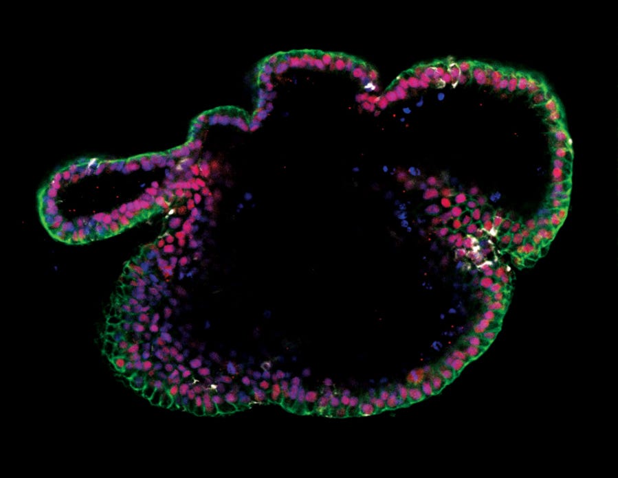
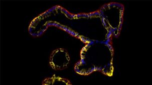
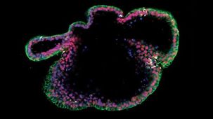
Immunocytochemistry image of an intestinal organoid cultured in mTeSR™ Plus and directed to intestinal organoids using the STEMdiff™ Intestinal Organoid Kit.

Figure 6. Generation of Intestinal Organoids from hPSCs Maintained in mTeSR™ Plus

Human ES (H9) cells were cultured with mTeSR™ Plus and directed to intestinal organoids using the STEMdiff™ Intestinal Organoid Kit. Image shows markers of the intestinal epithelium EpCAM (green) and CDX2 (red), and intestinal mesenchyme marker vimentin (white). Nuclei are counterstained with DAPI (blue).

产品说明书及文档

请在《产品说明书》中查找相关支持信息和使用说明,或浏览下方更多实验方案。

Document Type
Product Name
Catalog #
Lot #
Language
Catalog #
05140
Lot #
All
Language
English
Document Type
Technical Manual
Catalog #
05140
Lot #
All
Language
English
Document Type
Safety Data Sheet 1
Catalog #
05140
Lot #
All
Language
English
Document Type
Safety Data Sheet 2
Catalog #
05140
Lot #
All
Language
English
Document Type
Safety Data Sheet 3
Catalog #
05140
Lot #
All
Language
English
Document Type
Safety Data Sheet 4
Catalog #
05140
Lot #
All
Language
English
Document Type
Safety Data Sheet 5
Catalog #
05140
Lot #
All
Language
English
Document Type
Safety Data Sheet 6
Catalog #
05140
Lot #
All
Language
English

应用领域

本产品专为以下研究领域设计,适用于工作流程中的高亮阶段。探索这些工作流程,了解更多我们为各研究领域提供的其他配套产品。

相关材料与文献

技术资料 (49)

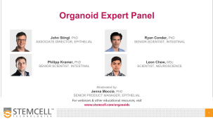
专家访谈

文献 (3)

Biallelic EPCAM deletions induce tissue-specific DNA repair deficiency and cancer predisposition V. J. Forster et al. NPJ Precision Oncology 2024 Mar

Abstract

We report a case of Mismatch Repair Deficiency (MMRD) caused by germline homozygous EPCAM deletion leading to tissue-specific loss of MSH2. Through the use of patient-derived cells and organoid technologies,we performed stepwise in vitro differentiation of colonic and brain organoids from reprogrammed EPCAM del iPSC derived from patient fibroblasts. Differentiation of iPSC to epithelial-colonic organoids exhibited continuous increased EPCAM expression and hypermethylation of the MSH2 promoter. This was associated with loss of MSH2 expression,increased mutational burden,MMRD signatures and MS-indel accumulation,the hallmarks of MMRD. In contrast,maturation into brain organoids and examination of blood and fibroblasts failed to show similar processes,preserving MMR proficiency. The combined use of iPSC,organoid technologies and functional genomics analyses highlights the potential of cutting-edge cellular and molecular analysis techniques to define processes controlling tumorigenesis and uncovers a new paradigm of tissue-specific MMRD,which affects the clinical management of these patients. Subject terms: Paediatric cancer,Cancer genetics
H and B Blood Antigens Are Essential for In Vitro Replication of GII.2 Human Norovirus S. Tamiya et al. Open Forum Infectious Diseases 2024 Dec

Abstract

Human norovirus (HuNoV) is a major cause of enteric infectious gastroenteritis and is classified into several genotypes based on its capsid protein amino acid sequence and nucleotide sequence of the polymerase gene. Among these,GII.4 is the major genotype worldwide. Epidemiological studies have highlighted the prevalence of GII.2. Although recent advances using human tissue– and induced pluripotent stem cell (iPSC)–derived intestinal epithelial cells (IECs) have enabled in vitro replication of multiple HuNoV genotypes,GII.2 HuNoV could replicate only in tissue-derived IECs and not in iPSC-derived IECs. We investigated the factors influencing GII.2 HuNoV replication in IECs,focusing on histo-blood group antigens. We also assessed the immunogenicity of GII.2 virus-like particles (VLPs) and their ability to induce neutralizing antibodies. Antibody cross-reactivity was tested to determine whether GII.2 VLPs could neutralize other HuNoV genotypes,including GII.4,GII.3,GII.6,and GII.17. Our findings indicated that GII.2 HuNoV replication in vitro requires the presence of both H and B antigens. Moreover,GII.2 VLPs generated neutralizing antibodies effective against both GII.2 and GII.4 but not against GII.3,GII.6,or GII.17. Comparatively,GII.2 and GII.17 VLPs induced broader neutralizing responses than GII.4 VLPs. The findings of this study suggests that GII.2 and GII.17 VLPs may be advantageous as HuNoV vaccine candidates because they elicit neutralizing antibodies against the predominant GII.4 genotype,which could be particularly beneficial for infants without prior HuNoV exposure. These insights will contribute to the development of effective HuNoV vaccines.
Organoid phenotypic screening identified glycyrrhizin that confers protection against tumor necrosis factor-induced cell death Y. Takahashi et al. Stem Cell Reports 2026 Apr

Abstract

Human organoids are considered physiological models that reflect human physiology; however,their applications in drug screening studies are limited. To develop a fundamental treatment for recurrent Crohn’s disease (CD),in which tumor necrosis factor (TNF) is a key pathogenic factor,we conducted phenotypic drug screening to prevent TNF-induced cell death in human intestinal epithelial organoids. Glycyrrhizin,a natural product of licorice root,dose-dependently blocked TNF-induced cell death in organoids but not in TNF-sensitive L929 cells; L929 cells exhibited necroptosis,whereas organoid-derived cells preferentially showed apoptosis upon TNF treatment,determining the specificity of glycyrrhizin. Glycyrrhizin inhibited downstream caspase-8 signaling,which is essential for TNF-dependent apoptosis,and ameliorated intestinal inflammation in vivo. These results demonstrate that glycyrrhizin may be a novel therapeutic compound for CD and highlight the importance of using organoids for phenotypic drug screening. Graphical abstract Highlights•Human intestinal organoids are sensitive to TNF-induced cytotoxicity•We developed an assay system to screen for compounds that resist TNF in organoids•Glycyrrhizin inhibited TNF-induced apoptosis but not necroptosis in organoids•Glycyrrhizin ameliorated intestinal inflammation in a murine model in vivo Takahashi et al. developed a high-throughput phenotypic screening platform using human intestinal organoids,which exhibited higher sensitivity to TNF than conventional intestinal epithelial cell lines,and identified glycyrrhizin that protected TNF-induced cell death both in organoids and in vivo. Phenotypic screening using organoids,as demonstrated in this study,paves the way for drug development and chemical biology.

更多信息

更多信息
物种
配方 无血清

法律声明:

STEMdiff™ Intestinal Organoid Kit was developed under license to intellectual property owned by the Cincinnati Children’s Hospital. 

质量保证:

产品仅供研究使用,不用于针对人或动物的诊断或治疗。 欲获悉更多关于STEMCELL的质控信息,请访问 STEMCELL.CN/COMPLIANCE.

Safety Statement:

CA WARNING: This product can expose you to Progesterone which is known to the State of California to cause cancer. For more information go to www.P65Warnings.ca.gov

Copyright © 2026 by STEMCELL Technologies. All rights reserved.

在线联系