若您需要咨询产品或有任何技术问题,请通过官方电话 400 885 9050 或邮箱 info.cn@stemcell.com 与我们联系。

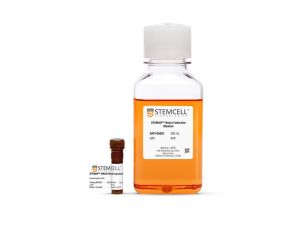
STEMdiff™ 前脑神经元成熟试剂盒

用于从人多能干细胞衍生的神经元前体细胞中生成功能性神经元的成熟试剂盒
只有 %1
¥4,024.00

产品号 #(选择产品)

产品号 #08605_C

用于从人多能干细胞衍生的神经元前体细胞中生成功能性神经元的成熟试剂盒

产品优势

  • 成分明确且无血清
  • 支持从 hPSC 衍生的神经元前体细胞高效生成功能性神经元
  • 生成高纯度的混合兴奋性和抑制性神经元群体(≥ 90% 的神经元),可能够在培养中长期维持
  • 针对使用 STEMdiff™ 前脑神经元分化试剂盒生成的神经元前体细胞的成熟进行了优化
  • 支持神经活动,以获得生理相关结果
  • 能够实现多个人胚胎干细胞和诱导多能干细胞系衍生的神经前体细胞的可重复成熟

产品组分包括

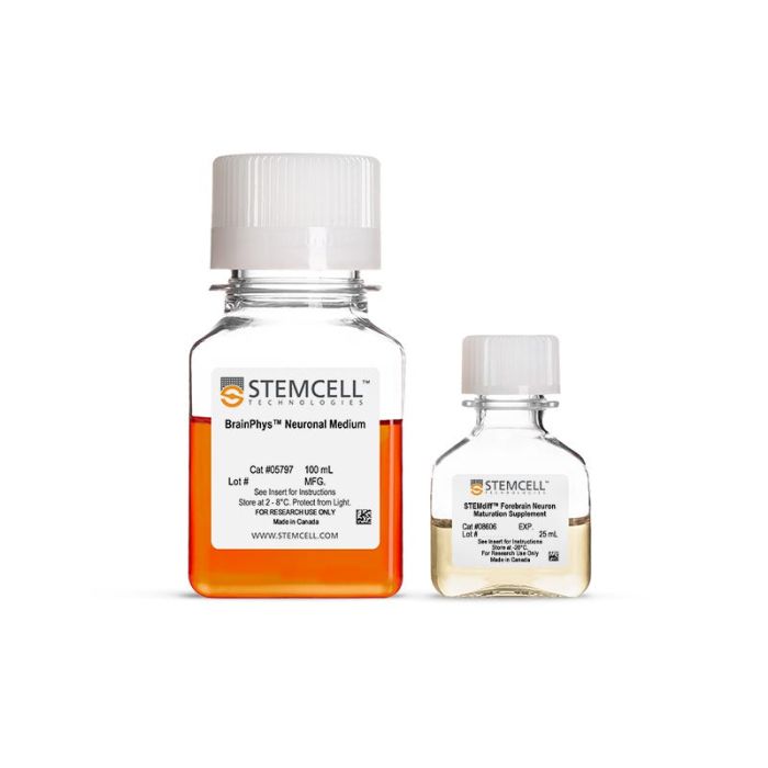
  • STEMdiff™ 前脑神经元成熟试剂盒,125 mL(产品号 #08605)
    • BrainPhys™ 神经元培养基,100 mL
    • STEMdiff™ 前脑神经元成熟添加物,25 mL
  • STEMdiff™ 前脑神经元成熟试剂盒,625 mL(产品号 #100-1659)
    • BrainPhys™ 神经元培养基,500 mL
    • STEMdiff™ 前脑神经元成熟添加物,125 mL

总览

STEMdiff™ 前脑神经元成熟试剂盒与 STEMdiff™ 前脑神经元分化试剂盒(产品号 #08600)配合使用,可从源自人多能干细胞的神经祖细胞中生成混合的前脑型(FOXG1 阳性)神经元群。分化所获得的神经元纯度高(III 类 β-微管蛋白阳性神经元≥ 90%;GFAP 阳性星形胶质细胞< 10%),功能齐全,可长期维持培养。使用这些产品获得的神经元是构建人神经发育和疾病模型、药物筛选、毒性检测和细胞疗法验证的多功能工具。


分类
专用培养基
 
细胞类型
神经细胞,PSC衍生,神经元
 
种属

 
应用
细胞培养,鉴定,分化,功能学筛选,免疫细胞化学,表型鉴定
 
品牌
STEMdiff
 
研究领域
疾病建模,药物发现和毒理检测,神经科学
 
制剂类别
无血清
 

实验数据

Figure 1. Schematic for the Embryoid Body Protocol

Forebrain-type neurons can be generated from NPCs after 6 days in STEMdiff™ Forebrain Neuron Differentiation Medium. For differentiation of precursors from embryonic and induced pluripotent stem cells, see the PIS. NPCs = neural progenitor cells; PIS = product information sheet

Figure 2. Schematic for the Monolayer Protocol

Forebrain-type neurons can be generated from NPCs after 6 days in STEMdiff™ Forebrain Neuron Differentiation Medium. For differentiation of precursors from embryonic and induced pluripotent stem cells, see the PIS. NPC = neural progenitor cells; PIS = product information sheet

Figure 3. Forebrain-Type Neurons Are Generated After Culture in STEMdiff™ Forebrain Neuron Differentiation and Maturation Kits

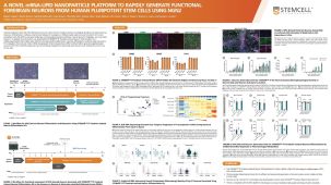
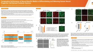
NPCs generated from hPSCs in mTeSR 1™ using the STEMdiff™ SMADi Neural Induction Kit EB protocol were differentiated and matured to forebrain-type neurons using the STEMdiff™ Forebrain Neuron Differentiation and Maturation Kits. (A) Forebrain-type neurons were formed after iPS cell-derived NPCs were cultured with the STEMdiff™ Forebrain Neuron Differentiation Kit for 7 days and STEMdiff™ Forebrain Neuron Maturation Kit for 14 days. The resulting cultures contain a highly pure population of (B) class III β-tubulin-positive neurons (green), with (C) fewer than 10% astrocytes (GFAP-positive cells, red). (D) Nuclei are labeled with DAPI (blue). NPCs = neural progenitor cells; hPSC = human pluripotent stem cell; EB = embryoid body; iPS = induced pluripotent stem

Figure 4. Downstream Differentiation of Neural Progenitor Cells to Neurons Is Possible Using the STEMdiff™ Differentiation and Maturation Kits

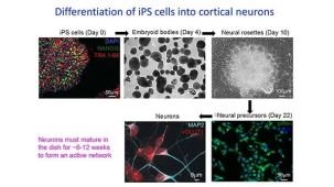
(A) NPCs generated from STiPS-R038 hPSCs in mTeSR™1 using the STEMdiff™ SMADi Neural Induction Kit EB protocol were differentiated and matured to cortical neurons using STEMdiff™ Forebrain Neuron Differentiation Kit for 7 days and STEMdiff™ Forebrain Neuron Maturation Kit for 14 days. The resulting cultures contain a highly pure population of (B) class III β-tubulin-positive neurons (green) with less than 10% GFAP-positive astrocytes (not shown). (C) The generated neurons are also positive for FOXG1 expression (red), indicating a forebrain-type identity. (D) Nuclei are labeled with Hoechst (blue). NPCs = neural progenitor cells; hPSC = human pluripotent stem cell

Figure 5. A Mixed Population of Forebrain-Type Cortical Neurons Is Generated Using the STEMdiff™ Differentiation and Maturation Kits

Forebrain-type neurons generated from iPSC-derived NPCs (line AIW002-02) were cultured using the STEMdiff™ Forebrain Neuron Differentiation Kit for 7 days and subsequently matured for the following 6 weeks using STEMdiff™ Forebrain Neuron Maturation Kit. The resulting cultures contain a mixed population of neurons expressing VGLUT1, a glutamatergic marker of excitatory neurons (green), as well as MAP2-positive neurons, indicating mature neurons (magenta). Nuclei are labeled with Hoechst (blue). Data courtesy of Cecilia Rocha, The Neuro's Early Drug Discovery Unit (EDDU), McGill University. iPSC = induced pluripotent stem cell; NPCs = neural progenitor cells

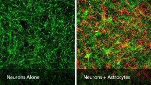
Figure 6. PSC-Derived Astrocytes and Neurons Can Be Co-Cultured to Model Cell-Cell Interactions In Vitro

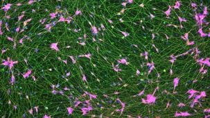
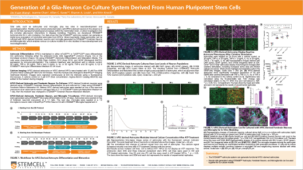
NPCs generated from the H1 cell line were differentiated to astrocytes using STEMdiff™ Astrocyte Differentiation and Maturation Kits. H9 cell-derived NPCs were differentiated to forebrain-type neurons using STEMdiff™ Forebrain Neuron Differentiation and Maturation Kits. For co-culture, matured astrocytes were seeded onto forebrain neurons that had been in STEMdiff™ Forebrain Neuron Maturation Medium for at least one week. Co-cultures were then switched to STEMdiff™ Forebrain Neuron Maturation Medium the following day and for the remaining co-culture. (A) Neurons cultured alone, following the co-culture feeding schedule, are labeled with DCX (green). (B) DCX-positive neurons (green) and astrocytes (GFAP, red) can be co-cultured for at least 1 - 2 weeks prior to analysis. For a detailed co-culture protocol, please see the Methods Library. NPCs = neural progenitor cells

Figure 7. PSC-Derived Neurons Survive and Mature when Co-Cultured with PSC-Derived Astrocytes

NPCs generated from the STiPS-R038 cell line were differentiated to astrocytes using STEMdiff™ Astrocyte Differentiation and Maturation Kits. STiPS-M001 cell-derived NPCs were differentiated to forebrain-type neurons using STEMdiff™ Forebrain Neuron Differentiation and Maturation Kits. After co-culture for one week, neurons (A) had significantly increased neurite outgrowth as measured on MAP2-positive neurons with the NeuriteTracer plugin for ImageJ (M Pool et al. J Neurosci Methods, 2008) and (B) were more numerous than neurons cultured alone using the same feeding schedule. *, p < 0.05; **, p < 0.01. NPCs = neural progenitor cells

产品说明书及文档

请在《产品说明书》中查找相关支持信息和使用说明,或浏览下方更多实验方案。

Document Type
Product Name
Catalog #
Lot #
Language
Catalog #
08605
Lot #
All
Language
English
Document Type
Safety Data Sheet
Catalog #
100-1659
Lot #
All
Language
English
Document Type
Safety Data Sheet 1
Catalog #
08605
Lot #
All
Language
English
Document Type
Safety Data Sheet 2
Catalog #
08605
Lot #
All
Language
English

应用领域

本产品专为以下研究领域设计,适用于工作流程中的高亮阶段。探索这些工作流程,了解更多我们为各研究领域提供的其他配套产品。

相关材料与文献

技术资料 (22)

文献 (6)

Human neural stem cell-derived artificial organelles to improve oxidative phosphorylation Nature Communications 2024 Sep

Abstract

Oxidative phosphorylation (OXPHOS) in the mitochondrial inner membrane is a therapeutic target in many diseases. Neural stem cells (NSCs) show progress in improving mitochondrial dysfunction in the central nervous system (CNS). However,translating neural stem cell-based therapies to the clinic is challenged by uncontrollable biological variability or heterogeneity,hindering uniform clinical safety and efficacy evaluations. We propose a systematic top-down design based on membrane self-assembly to develop neural stem cell-derived oxidative phosphorylating artificial organelles (SAOs) for targeting the central nervous system as an alternative to NSCs. We construct human conditionally immortal clone neural stem cells (iNSCs) as parent cells and use a streamlined closed operation system to prepare neural stem cell-derived highly homogenous oxidative phosphorylating artificial organelles. These artificial organelles act as biomimetic organelles to mimic respiration chain function and perform oxidative phosphorylation,thus improving ATP synthesis deficiency and rectifying excessive mitochondrial reactive oxygen species production. Conclusively,we provide a framework for a generalizable manufacturing procedure that opens promising prospects for disease treatment. Regulating oxidative phosphorylation and restoring redox homeostasis are crucial in neurological disorders. Here,the authors develop a top-down membrane self-assembly strategy to develop stem cell-derived artificial organelles (SAOs) that mimic mitochondrial oxidative phosphorylation without the risks associated with stem cell therapy.
GRAMD1B is a regulator of lipid homeostasis, autophagic flux and phosphorylated tau D. A. Ingram et al. Nature Communications 2025 Apr

Abstract

Lipid dyshomeostasis and tau pathology are present in frontotemporal lobar degeneration (FTLD) and Alzheimer’s disease (AD). However,the relationship between lipid dyshomeostasis and tau pathology remains unclear. We report that GRAM Domain Containing 1B (GRAMD1B),a nonvesicular cholesterol transporter,is increased in excitatory neurons of human neural organoids (HNOs) with the MAPT R406W mutation. Human FTLD,AD cases,and PS19 tau mice also have increased GRAMD1B expression. We show that overexpression of GRAMD1B increases levels of free cholesterol,lipid droplets,and impairs autophagy flux. Modulating GRAMD1B in iPSC-derived neurons also alters key autophagy-related components such as PI3K,phospho-AKT,and p62,as well as phosphorylated tau,and CDK5R1. Blocking GRAMD1B function decreases free cholesterol and lipid droplets. Knocking down GRAMD1B additionally reduces phosphorylated tau,and CDK5R1 expression. Our findings elucidate the role of GRAMD1B in the nervous system and highlight its relevance to FTLD and AD. Subject terms: Diseases of the nervous system,Ageing
AAV vectors trigger DNA damage response-dependent pro-inflammatory signalling in human iPSC-derived CNS models and mouse brain H. Costa-Verdera et al. Nature Communications 2025 Apr

Abstract

Adeno-associated viral (AAV) vector-based gene therapy is gaining foothold as treatment for genetic neurological diseases with encouraging clinical results. Nonetheless,dose-dependent adverse events have emerged in recent clinical trials through mechanisms that remain unclear. We have modelled here the impact of AAV transduction in cell models of the human central nervous system (CNS),taking advantage of induced pluripotent stem cells. Our work uncovers vector-induced innate immune mechanisms that contribute to cell death. While empty AAV capsids were well tolerated,the AAV genome triggered p53-dependent DNA damage responses across CNS cell types followed by the induction of inflammatory responses. In addition,transgene expression led to MAVS-dependent activation of type I interferon responses. Formation of DNA damage foci in neurons and gliosis were confirmed in murine striatum upon intraparenchymal AAV injection. Transduction-induced cell death and gliosis could be prevented by inhibiting p53 or by acting downstream on STING- or IL-1R-mediated responses. Together,our work identifies innate immune mechanisms of vector sensing in the CNS that can potentially contribute to AAV-associated neurotoxicity. Subject terms: Neuroimmunology,Innate immunity,Neural stem cells

更多信息

更多信息
物种
配方 无血清
质量保证:

产品仅供研究使用,不用于针对人或动物的诊断或治疗。 欲获悉更多关于STEMCELL的质控信息,请访问 STEMCELL.CN/COMPLIANCE.
Copyright © 2026 by STEMCELL Technologies. All rights reserved.

在线联系