若您需要咨询产品或有任何技术问题,请通过官方电话 400 885 9050 或邮箱 info.cn@stemcell.com 与我们联系。

STEMdiff™ 心室肌细胞分化试剂盒

用于将人 PSC 分化为心室肌细胞以及长期维持人PSC衍生心肌细胞的无血清培养基
只有 %1
¥11,062.00

产品号 #(选择产品)

产品号 #05010_C

用于将人 PSC 分化为心室肌细胞以及长期维持人PSC衍生心肌细胞的无血清培养基

产品优势

  • 支持整个hPSC衍生心肌细胞工作流程
  • 简单的单层方案可在 15 天内获得心肌细胞
  • 一套无血清试剂盒可产生超过 5000 万个心肌细胞 (cTnT+)
  • 在多种人PSC细胞系中表现稳定,变异性小

产品组分包括

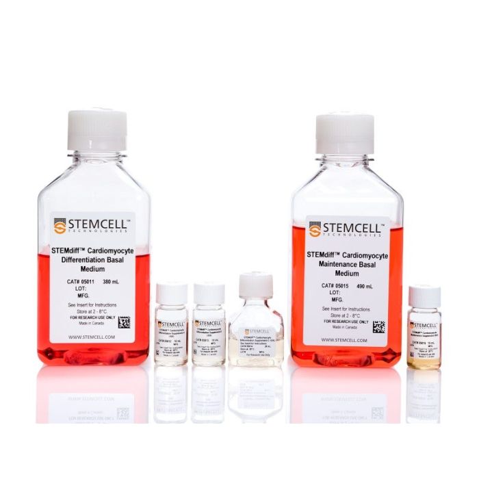
  • STEMdiff™ 心肌细胞分化基础培养基,380 mL
  • STEMdiff™ 心室肌细胞分化补充剂 A (10X),10 mL
  • STEMdiff™ 心室肌细胞分化补充剂 B (10X),10 mL
  • STEMdiff™ 心室肌细胞分化补充剂 C (10X),20 mL
  • STEMdiff™ 心肌细胞维持基础培养基,490 mL
  • STEMdiff™ 心肌细胞维持补充剂 (50X),10 mL
Need a high-quality cell source? Use the hiPSC SCTi003-A (female) or SCTi004-A (male) control lines, manufactured with mTeSR™ Plus.
专为您的实验方案打造的产品
要查看实验方案所需的所有配套产品,请参阅《实验方案与技术文档》

总览

STEMdiff™ 心室肌细胞分化试剂盒(产品号 #05010)包含用于将人胚胎干细胞(ES)和诱导多能干细胞(iPS)(统称为人多能干细胞 [hPSCs])分化为心室肌细胞(肌钙蛋白T阳性 [cTnT+])的培养基,以及用于维持 hPSC 衍生心肌细胞的培养基。该试剂盒为无血清体系,可用于从以mTeSR™1(产品号 #85850)、mTeSR™ Plus(产品号 #100-0276)、TeSR™-AOF(产品号 #100-0401)或TeSR™-E8™(产品号 #05990)培养的hPSC团块中诱导分化出心室肌细胞。这些细胞中超过80%为cTnT+。平均每12孔板的单孔可收获1 x 10^6个细胞。

STEMdiff™ 心肌细胞维持试剂盒(产品号 #05020)包含维持基础培养基和补充剂,可用于长期维持hPSC衍生的心肌细胞一个月或更长时间。这些心肌细胞可用于各种下游应用和分析。

注:本产品原名为“STEMdiff™ 心肌细胞分化试剂盒”;产品本身和生产流程未发生变化,但名称已更新,以更准确地反映生成的细胞类型。


分类
专用培养基
 
细胞类型
心肌细胞,PSC衍生
 
种属

 
应用
细胞培养,分化,培养
 
品牌
STEMdiff
 
研究领域
疾病建模,药物发现和毒理检测,干细胞生物学
 
制剂类别
无血清
 

实验数据

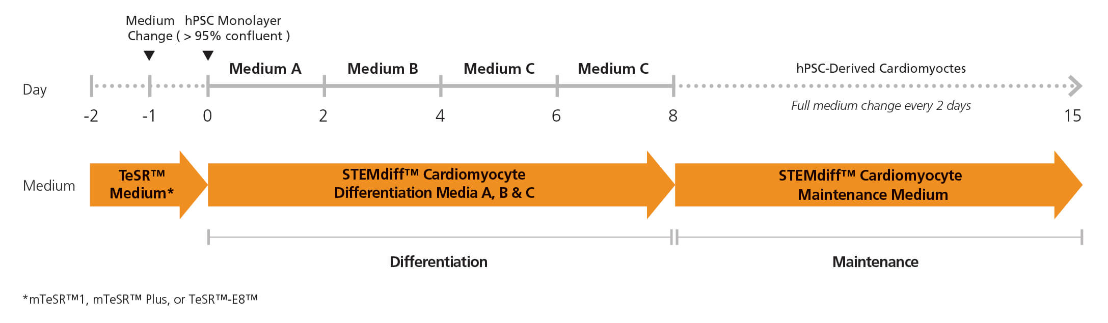
Figure 1. Cardiomyocyte Differentiation Protocol

Two days before the differentiation protocol, hPSC colonies are harvested and seeded as single cells at 350,000 cells/well in a 12-well format in TeSR™ medium. After one day (Day -1), the medium is replaced with fresh TeSR™ medium. The following day (Day 0), the TeSR™ medium is replaced with Medium A (STEMdiff™ Cardiomyocyte Differentiation Basal Medium containing Supplement A) to begin inducing the cells toward a cardiomyocyte fate. On day 2, a full medium change is performed with fresh Medium B (STEMdiff™ Cardiomyocyte Differentiation Basal Medium containing Supplement B). On days 4 and 6, full medium changes are performed with fresh Medium C (STEMdiff™ Cardiomyocyte Differentiation Basal Medium containing Supplement C). On day 8, medium is switched to STEMdiff™ Cardiomyocyte Maintenance Medium with full medium changes on days 10, 12 and 14, to promote further differentiation into cardiomyocyte cells. Small beating areas of cardiomyocytes can be seen as early as day 8, progressing to a full lawn of beating cardiomyocytes that can be harvested as early as day 15.

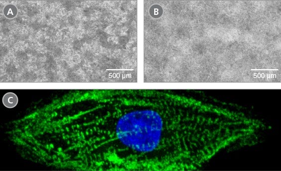
Figure 2. Morphology of hPSC-Derived Cardiomyocytes

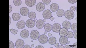
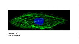
Representative images of (A) hES (H9) cells and (B) hiPS (WLS-1C) cells on day 15 of differentiation to cardiomyocytes using the STEMdiff™ Ventricular Cardiomyocyte Differentiation Kit. Differentiated cells exhibit typical cardiomyocyte morphology as an adherent, tightly packed web-like monolayer of beating cells. (C) Representative confocal microscopy image of a single hPSC-derived cardiomyocyte generated with the STEMdiff™ Ventricular Cardiomyocyte Differentiation Kit and stained with cTnT (green) and DAPI (blue).

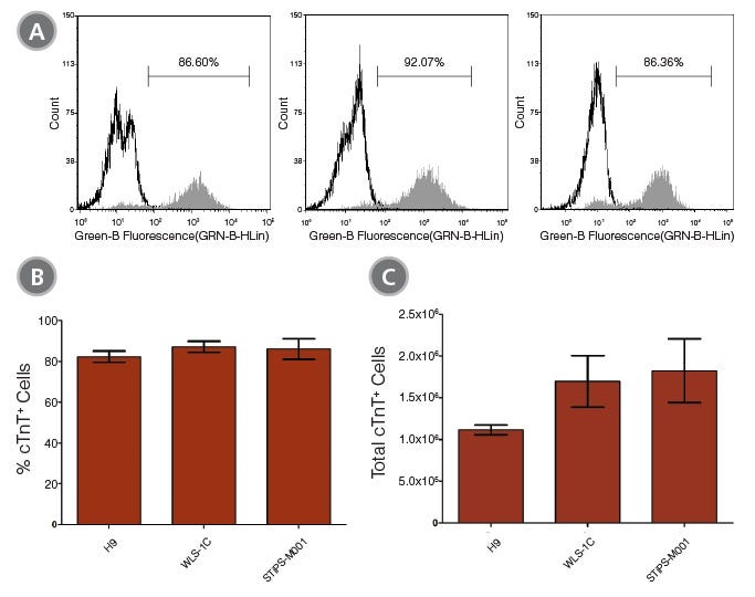
Figure 3. Efficient and Robust Generation of cTnT-Positive Cardiomyocytes

hES and hiPS cells were cultured for 15 days in single wells of 12-well plates using the STEMdiff™ Ventricular Cardiomyocyte Differentiation Kit. At the end of the culture period, cells were harvested and analyzed by flow cytometry for expression of cardiac troponin T (cTnT). (A) Histogram analysis for cardiomyocyte cell marker cTnT for cultures of hES (H9) and hiPS (WLS-1C and STiPS-M001) cells. (Filled = sample; blank = secondary antibody only control) (B,C) Percentages and total numbers of cells expressing cTnT in cultures of hES or hiPS cells are shown. Data shown as mean ± SEM; n=3.

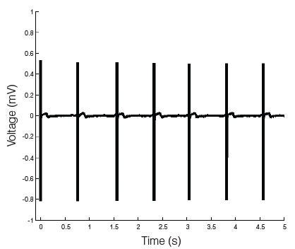
Figure 4. hPSC-Derived Cardiomyocytes Exhibit a Robust and Stable Excitability Profile

Microelectrode array (MEA) voltage recordings of cardiomyocytes (day 27) derived from human pluripotent stem cells generated and maintained with the STEMdiff™ Cardiomyocyte Differentiation and Maintenance Kits. The hPSC-derived cardiomyocytes have a characteristic electrical profile and stable beat rate. A large depolarization spike followed by a smaller repolarization deflection is observed.

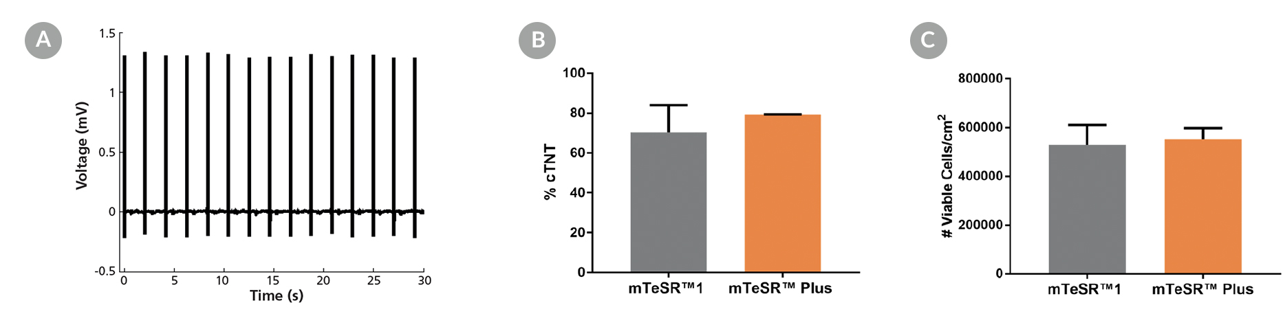
Microelectrode array and flow cytometry of human ES and iPS cells maintained in mTeSR™1 (daily feeds) or mTeSR™ Plus (restricted feeds) and differentiated to cardiomyocytes using the STEMdiff™ Ventricular Cardiomyocyte Differentiation Kit.

Figure 5. Generation of Cardiomyocytes from hPSCs Maintained in mTeSR™ Plus

Human ES (H9) and iPS (WLS-1C) cells were maintained in mTeSR™1 (daily feeds) or mTeSR™ Plus (restricted feeds) and differentiated to cardiomyocytes using the STEMdiff™ Ventricular Cardiomyocyte Differentiation Kit. At the end of the differentiation period, cells were harvested and analyzed by microelectrode array (MEA) and flow cytometry. (A) Representative MEA voltage recordings of cardiomyocytes (day 20) demonstrate a characteristic electrical profile and stable beat rate. (B) Percentages of cells expressing cTNT and (C) total number of viable cells harvested are shown. Data are expressed as the mean (± SEM); n=2.

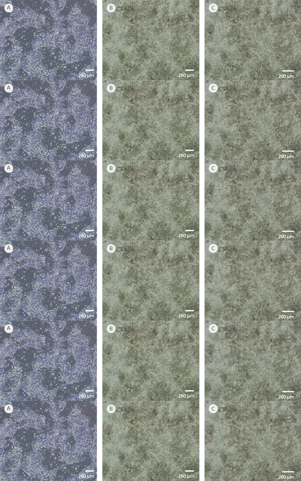
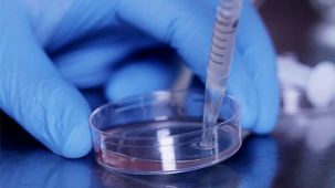
Microscopy images of iPSCdirect cells and differentiated ventral cardiomyocytes, and a video of coordinated contraction or beating behavior of cardiomyocytes in a culture dish

Figure 6. iPSCdirect™ SCTi003-A Human Pluripotent Stem Cells Can Successfully Differentiate into Ventricular Cardiomyocytes

Ventricular cardiomyocytes were generated from iPSCdirect™ SCTi003-A cells using STEMdiff™ Ventricular Cardiomyocyte Differentiation Kit (Catalog #05010). (A) 48 hours after thawing and plating in mTeSR™ Plus and CloneR™2, iPSCdirect™ cells reached the desired confluency and are ready for Day 0 of differentiation according to the STEMdiff™ Ventricular Cardiomyocyte Product Information Sheet. (B) By Day 15 of differentiation, monolayer cultures show iPSC-derived ventricular cardiomyocytes that (C) exhibit coordinated beating behavior.

产品说明书及文档

请在《产品说明书》中查找相关支持信息和使用说明,或浏览下方更多实验方案。

Document Type
Product Name
Catalog #
Lot #
Language
Catalog #
05010
Lot #
All
Language
English
Document Type
Safety Data Sheet 1
Catalog #
05010
Lot #
All
Language
English
Document Type
Safety Data Sheet 2
Catalog #
05010
Lot #
All
Language
English
Document Type
Safety Data Sheet 3
Catalog #
05010
Lot #
All
Language
English
Document Type
Safety Data Sheet 4
Catalog #
05010
Lot #
All
Language
English
Document Type
Safety Data Sheet 5
Catalog #
05010
Lot #
All
Language
English
Document Type
Safety Data Sheet 6
Catalog #
05010
Lot #
All
Language
English

应用领域

本产品专为以下研究领域设计,适用于工作流程中的高亮阶段。探索这些工作流程,了解更多我们为各研究领域提供的其他配套产品。

相关材料与文献

技术资料 (17)

文献 (16)

Ex Vivo Expansion of CD34+ CD90+ CD49f+ Hematopoietic Stem and Progenitor Cells from Non-Enriched Umbilical Cord Blood with Azole Compounds. S. Bari et al. Stem cells translational medicine 2018

Abstract

Umbilical cord blood (UCB) transplants in adults have slower hematopoietic recovery compared to bone marrow (BM) or peripheral blood (PB) stem cells mainly due to low number of total nucleated cells and hematopoietic stem and progenitor cells (HSPC). As such in this study,we aimed to perform ex vivo expansion of UCB HSPC from non-enriched mononucleated cells (MNC) using novel azole-based small molecules. Freshly-thawed UCB-MNC were cultured in expansion medium supplemented with small molecules and basal cytokine cocktail. The effects of the expansion protocol were measured based on in vitro and in vivo assays. The proprietary library of {\textgreater}50 small molecules were developed using structure-activity-relationship studies of SB203580,a known p38-MAPK inhibitor. A particular analog,C7,resulted in 1,554.1 ± 27.8-fold increase of absolute viable CD45+ CD34+ CD38- CD45RA- progenitors which was at least 3.7-fold higher than control cultures (p {\textless} .001). In depth phenotypic analysis revealed {\textgreater}600-fold expansion of CD34+ /CD90+ /CD49f+ rare HSPCs coupled with significant (p {\textless} .01) increase of functional colonies from C7 treated cells. Transplantation of C7 expanded UCB grafts to immunodeficient mice resulted in significantly (p {\textless} .001) higher engraftment of human CD45+ and CD45+ CD34+ cells in the PB and BM by day 21 compared to non-expanded and cytokine expanded grafts. The C7 expanded grafts maintained long-term human multilineage chimerism in the BM of primary recipients with sustained human CD45 cell engraftment in secondary recipients. In conclusion,a small molecule,C7,could allow for clinical development of expanded UCB grafts without pre-culture stem cell enrichment that maintains in vitro and in vivo functionality. Stem Cells Translational Medicine 2018;7:376-393.
Linking a cell-division gene and a suicide gene to define and improve cell therapy safety. Q. Liang et al. Nature 2018

Abstract

Human pluripotent cell lines hold enormous promise for the development of cell-based therapies. Safety,however,is a crucial prerequisite condition for clinical applications. Numerous groups have attempted to eliminate potentially harmful cells through the use of suicide genes1,but none has quantitatively defined the safety level of transplant therapies. Here,using genome-engineering strategies,we demonstrate the protection of a suicide system from inactivation in dividing cells. We created a transcriptional link between the suicide gene herpes simplex virus thymidine kinase (HSV-TK) and a cell-division gene (CDK1); this combination is designated the safe-cell system. Furthermore,we used a mathematical model to quantify the safety level of the cell therapy as a function of the number of cells that is needed for the therapy and the type of genome editing that is performed. Even with the highly conservative estimates described here,we anticipate that our solution will rapidly accelerate the entry of cell-based medicine into the clinic.
Development of induced pluripotent stem cells from a patient with hypertrophic cardiomyopathy who carries the pathogenic myosin heavy chain 7 mutation p.Arg403Gln. M. Holliday et al. Stem cell research 2018

Abstract

Hypertrophic cardiomyopathy (HCM) is an inherited cardiomyopathy characterized by left ventricular hypertrophy ≥15 mm in the absence of loading conditions. HCM has a prevalence of up to one in 200,and can result in significant adverse outcomes including heart failure and sudden cardiac death. An induced pluripotent stem cell (iPSC) line was generated from peripheral blood mononuclear cells obtained from the whole blood of a 38-year-old female patient with HCM in which genetic testing identified the well-known pathogenic p.Arg403Gln mutation in myosin heavy chain 7. iPSCs express pluripotency markers,demonstrate trilineage differentiation capacity,and display a normal 46,XX female karyotype. This resource will allow further assessment of the pathophysiological development of HCM.

更多信息

更多信息
物种
配方 无血清

质量保证:
本产品仅供科研使用,除非另有说明,不得用于人体或动物的诊断或治疗用途。如需了解 STEMCELL 的质量体系,请访问www.stemcell.cn/compliance

Copyright © 2026 by STEMCELL Technologies. All rights reserved.

在线联系