若您需要咨询产品或有任何技术问题,请通过官方电话 400 885 9050 或邮箱 info.cn@stemcell.com 与我们联系。

STEMdiff™ 星形胶质细胞分化试剂盒

无血清分化试剂盒,用于从人多能干细胞(hPSC)来源的神经前体细胞生成星形胶质细胞前体。
只有 %1
¥4,264.00

产品号 #(选择产品)

产品号 #100-0013_C

无血清分化试剂盒,用于从人多能干细胞(hPSC)来源的神经前体细胞生成星形胶质细胞前体。

产品优势

  • 成分明确且无血清
  • 支持高效生成具有功能性的星形胶质细胞
  • 针对使用 STEMdiff™ SMADi 神经诱导试剂盒生成的神经前体细胞进行优化分化
  • 可重复地从多种人胚胎干细胞(ES)和诱导多能干细胞(iPS)系中生成皮层类型的星形胶质细胞前体

产品组分包括

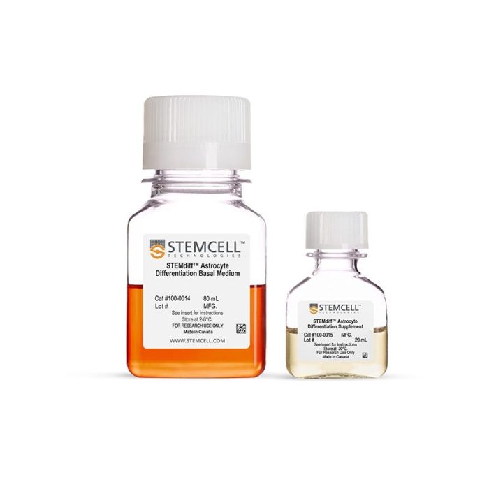
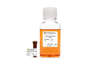
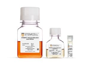
  • STEMdiff™ 星形胶质细胞分化基础培养基,80 mL
  • STEMdiff™ 星形胶质细胞分化补充剂,20 mL
Need a high-quality cell source? Use the hiPSC SCTi003-A (female) or SCTi004-A (male) control lines, manufactured with mTeSR™ Plus.

总览

STEMdiff™ 星形胶质细胞分化试剂盒可用于快速且高效地从使用 STEMdiff™ SMADi 神经诱导试剂盒(目录号:08581)获得的人多能干细胞(hPSC)来源的神经前体细胞(NPCs)中生成星形胶质细胞前体。随后,这些星形胶质细胞前体可通过 STEMdiff™ 无血清星形胶质细胞成熟试剂盒(目录号:100-1666)进一步成熟为星形胶质细胞。

采用该无血清系统,最短仅需 7 周即可从 hPSC 生成高度纯化的星形胶质细胞群体(平均 >70% S100B 阳性、>60% GFAP 阳性星形胶质细胞,<15% doublecortin 阳性神经元),且这些细胞可在体外长期培养维持。使用该系列产品衍生的细胞可广泛应用于人类神经发育和疾病模型构建、药物筛选、毒性测试以及细胞治疗验证等研究领域。

分类
专用培养基
 
细胞类型
神经细胞,PSC衍生,神经干/祖细胞
 
种属

 
应用
细胞培养,分化
 
品牌
STEMdiff
 
研究领域
疾病建模,药物发现和毒理检测,神经科学
 
制剂类别
无血清
 

实验数据

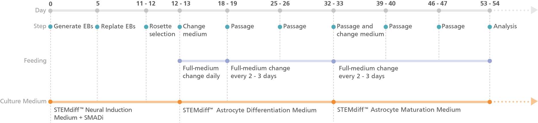
Experimental Protocol Schematic for STEMdiff™ Forebrain Neuron Differentiation and Maturation Kits (Embryoid Body Protocol)

Figure 1. Schematic for the Embryoid Body Protocol

Cortical-type astrocyte precursors can be generated in 20 days from hPSC-derived neural progenitor cells (NPCs) after selecting neural rosettes from replated embryoid bodies. For the maturation of precursors to cortical-type astrocytes, see the PIS.

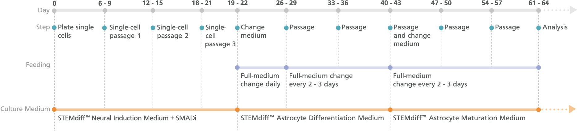
Experimental Protocol Schematic for STEMdiff™ Forebrain Neuron Differentiation and Maturation Kits (Monolayer Protocol)

Figure 2. Schematic for the Monolayer Protocol

Cortical-type astrocyte precursors can be generated in 21 days from neural progenitor cell (NPC) monolayers derived from embryonic and induced pluripotent stem cells after three single-cell passages. For the maturation of precursors to cortical-type astrocytes, see the PIS.

Culturing PSCs in STEMdiff™ SMADi Neural Induction Kit and STEMdiff™ Astrocyte Differentiation and Maturation Kits Yields Cortical-Type Astrocytes

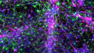
Figure 3. Cortical-Type Astrocytes Are Generated After Culture in STEMdiff™ Astrocyte Differentiation and Maturation Kits

NPCs generated from hPSCs in TeSR™-E8™ using the STEMdiff™ SMADi Neural Induction Kit embryoid body (EB) protocol were differentiated and matured to cortical-type astrocytes using the STEMdiff™ Astrocyte Differentiation and Maturation Kits. Cortical-type astrocytes were formed after iPS cell-derived NPCs were cultured with the STEMdiff™ Astrocyte Differentiation Kit for 3 weeks and STEMdiff™ Astrocyte Maturation Kit for 3 weeks. (A) Nuclei are labeled with DAPI (gray). The resulting cultures contain a highly pure population of astrocytes, which are (B) more than 60% GFAP-positive (green) and (C) more than 70% S100B-positive (magenta), with (D) fewer than 15% neurons (DCX-positive cells, cyan). Scale bar = 100 μm.

Figure 4. STEMdiff™ Astrocyte Kits Generate Cells Expressing Expected Levels of Genes Characteristic for Astrocytes

Embryonic stem and induced pluripotent stem cells from a variety of lines (n = 6, maintained in mTeSR™1 or TeSR™-E8™) were differentiated to NPCs using the STEMdiff™ SMADi Neural Induction Kit embryoid body protocol. Cells were then grown in STEMdiff™ Astrocyte Differentiation Kit for 3 weeks followed by STEMdiff™ Astrocyte Maturation Kit for 3 weeks prior to analysis. Expression levels were measured by quantitative PCR (qPCR) and normalized to hPSC controls relative to housekeeping genes 18S and TBP.

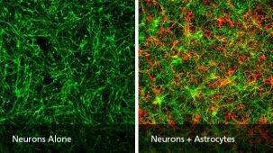
Figure 5. PSC-Derived Astrocytes and Neurons Can Be Co-Cultured to Model Cell-Cell Interactions In Vitro

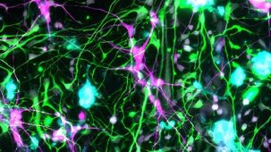
NPCs generated from the H1 cell line were differentiated to astrocytes using STEMdiff™ Astrocyte Differentiation and Maturation Kits. H9 cell-derived NPCs were differentiated to forebrain-type neurons using STEMdiff™ Forebrain Neuron Differentiation and Maturation Kits. For co-culture, matured astrocytes were seeded onto forebrain neurons that had been in STEMdiff™ Forebrain Neuron Maturation Medium for at least one week. Co-cultures were then switched to STEMdiff™ Forebrain Neuron Maturation Medium the following day and for the remaining co-culture. (A) Neurons cultured alone, following the co-culture feeding schedule, are labeled with DCX (green). (B) DCX-positive neurons (green) and astrocytes (GFAP, red) can be co-cultured for at least 1 - 2 weeks prior to analysis. For a detailed co-culture protocol, please see the Methods Library.

Figure 6. PSC-Derived Neurons Survive and Mature when Co-Cultured with PSC-Derived Astrocytes

NPCs generated from the STiPS-R038 cell line were differentiated to astrocytes using STEMdiff™ Astrocyte Differentiation and Maturation Kits. STiPS-M001 cell-derived NPCs were differentiated to forebrain-type neurons using STEMdiff™ Forebrain Neuron Differentiation and Maturation Kits. After co-culture for one week, neurons (A) had significantly increased neurite outgrowth as measured on MAP2-positive neurons with the NeuriteTracer plugin for ImageJ (M Pool et al. J Neurosci Methods, 2008) and (B) were more numerous than neurons cultured alone using the same feeding schedule. *, p < 0.05

产品说明书及文档

请在《产品说明书》中查找相关支持信息和使用说明,或浏览下方更多实验方案。

Document Type
Product Name
Catalog #
Lot #
Language
Catalog #
100-0013
Lot #
All
Language
English
Document Type
Safety Data Sheet 1
Catalog #
100-0013
Lot #
All
Language
English
Document Type
Safety Data Sheet 2
Catalog #
100-0013
Lot #
All
Language
English

应用领域

本产品专为以下研究领域设计,适用于工作流程中的高亮阶段。探索这些工作流程,了解更多我们为各研究领域提供的其他配套产品。

相关材料与文献

技术资料 (30)

文献 (3)

The Alzheimer's disease‐associated complement receptor 1 variant confers risk by impacting glial phagocytosis N. Daskoulidou et al. Alzheimer's & Dementia 2025 Jul

Abstract

Genome‐wide association studies have implicated complement in Alzheimer's disease (AD). The CR1*2 variant of complement receptor 1 (CR1; CD35),confers increased AD risk. We confirmed CR1 expression on glial cells; however,how CR1 variants influence AD risk remains unclear. Induced pluripotent stem cell‐derived microglia and astrocytes were generated from donors homozygous for the common CR1 variants (CR1*1/CR1*1;CR1*2/CR1*2). CR1 expression was quantified and phagocytic activity assessed using diverse targets ( Escherichia coli bioparticles,amyloid β aggregates,and synaptoneurosomes),with or without serum opsonization. Expression of CR1*1 was significantly higher than CR1*2 on glial lines. Phagocytosis for all targets was markedly enhanced following serum opsonization,attenuated by Factor I‐depletion,demonstrating CR1 requirement for C3b processing. CR1*2‐expressing glia showed significantly enhanced phagocytosis of all opsonized targets compared to CR1*1‐expressing cells. CR1 is critical for glial phagocytosis of opsonized targets. CR1*2,despite lower expression,enhances glial phagocytosis,providing mechanistic explanation of increased AD risk. Induced pluripotent stem cell (iPSC)‐derived glia from individuals expressing the Alzheimer's disease (AD) risk variant complement receptor (CR) 1*2 exhibit lower CR1 expression compared to those from donors expressing the non‐risk form CR1*1. The iPSC‐derived glia from individuals expressing the AD risk variant CR1*2 exhibit enhanced phagocytic activity for opsonized bacterial particles,amyloid‐β aggregates and human synaptoneurosomes compared to those from donors expressing the non‐risk form CR1*1. We suggest that expression of the CR1*2 variant confers risk of AD by enhancing the phagocytic capacity of glia for opsonized targets.
MicroRNA‐153‐3p targets repressor element 1‐silencing transcription factor (REST) and neuronal differentiation: Implications for Alzheimer's disease R. Wang et al. Alzheimer's & Dementia 2025 Aug

Abstract

Small non‐coding microRNAs (miRNAs) play essential roles in Alzheimer's disease (AD) pathogenesis. Repressor element 1‐silencing transcription factor (REST) is involved in AD,though its regulation remains unclear. We performed real‐time quantitative polymerase chain reaction (qPCR) in autopsied brain tissues to determine miR‐153‐3p and AD associations. A reporter‐based assay measured the activity of REST mRNA 3′‐untranslated region (3′‐UTR). Induced pluripotent stem cells (iPSC)‐derived neurons and human cell lines were applied to determine miR‐153‐3p regulation of endogenous proteins. Elevation of miR‐153‐3p is associated with a reduced probability of AD,while elevated REST is associated with a greater probability of AD. The 3′‐UTR functional assay pinpointed the miR‐153‐3p binding sites. miR‐153‐3p treatment reduced REST,amyloid precursor protein (APP),and α‐synuclein (SNCA) 3′‐UTR activities and protein levels. miR‐153‐3p treatment altered REST and neuronal differentiation in iPSC‐derived neuronal stem cells. RNA‐sequencing and proteomics revealed miR‐153‐3p‐associated networks. miR‐153‐3p reduces REST,APP,and SNCA expression,pointing toward its therapeutic and biomarker potential in neurodegenerative diseases. With the increased emphasis on comorbidities of Alzheimer's disease (AD) and other neurodegenerative diseases,we identified that miR‐153‐3p,as a master regulator,reduced a group of neurodegeneration related proteins: REST,amyloid precursor protein (APP) and α‐synuclein (SNCA) levels. The elevation of miR‐153‐3p levels is associated with reduced probability of AD in posterior cingulate cortex (PCC),while REST,by contrast,is associated with a greater probability of AD. miR‐153‐3p treatment alters REST protein levels and neuronal differentiation in induced pluripotent stem cells (iPSC) derived neuronal cells. RNA sequencing proteomics and interactome analysis revealed the role of miR‐153‐3p in axonal guidance.
Modeling neurovascular dysfunction in Alzheimer’s disease using an isogenic brain-chip model A. N. Shen et al. Fluids and Barriers of the CNS 2026 Jan

Abstract

Background: The pathology of Alzheimer’s Disease (AD) is characterized by aggregates of amyloid beta (Aβ) peptides and neurofibrillary tau tangles. Increased blood-brain barrier (BBB) permeability and reduced Aβ clearance,which signal neurovascular dysfunction,have also been proposed as early markers of AD. Despite intense scrutiny,the mechanisms of AD remain elusive and novel treatments that address core symptoms of dementia are limited. New alternative methods (NAMs) aim to develop in-vitro translational models that recapitulate human pathology more accurately than previous models and could contribute to the development of new therapies. Methods: Here,we developed a NAM model of the cortical neurovascular unit (NVU) using brain cells derived from human induced pluripotent stem cells (hiPSCs) from a patient with AD and a healthy individual. Differentiated neurons,astrocytes,pericytes,microglia,and brain-like microvascular endothelial cells were cultured in a microphysiological system to create a brain-chip model to evaluate NVU-related endpoints. Results: Compared to control,AD brain-chips had reduced claudin-5 and ZO-1 expression and increased paracellular permeability. AD brain-chips also had decreased activity of the efflux transporter P-glycoprotein (P-gp),but its expression was unchanged. In AD brain-chips,levels of Aβ42,total tau,and p-tau 181 were decreased in protein lysates from the brain channel,while levels of total tau and p-tau 181 were increased in protein lysates from the vascular channel. Finally,AD brain-chips had increased levels of the proinflammatory markers IL-6 and MCP-1 in effluent from both brain and vascular channels. Conclusion: In this brain-chip model,we showed Aβ-independent NVU dysfunction that was related to neuroinflammation and vascular tau accumulation. This study demonstrates the utility of the brain-chip model to evaluate changes in NVU functions induced by AD-like pathology and highlights donor-specific responses associated with the use of hiPSC-derived models.

更多信息

更多信息
物种
配方 无血清
质量保证:

产品仅供研究使用,不用于针对人或动物的诊断或治疗。 欲获悉更多关于STEMCELL的质控信息,请访问 STEMCELL.CN/COMPLIANCE.
Copyright © 2026 by STEMCELL Technologies. All rights reserved.

在线联系