若您需要咨询产品或有任何技术问题,请通过官方电话 400 885 9050 或邮箱 info.cn@stemcell.com 与我们联系。

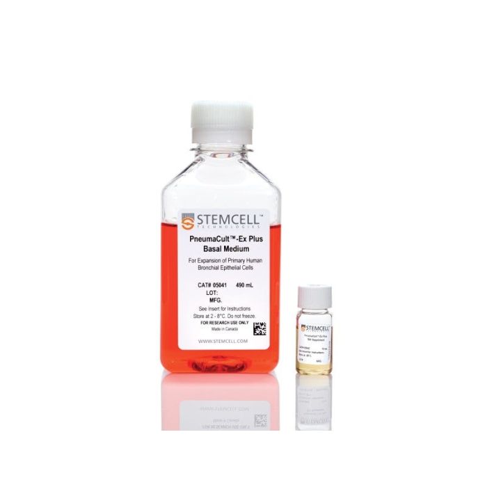
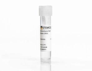
PneumaCult™-Ex Plus 培养基

用于扩增人原代呼吸道上皮细胞的无血清和无BPE培养基
只有 %1
¥3,498.00

产品号 #(选择产品)

产品号 #05040

用于扩增人原代呼吸道上皮细胞的无血清和无BPE培养基

产品优势

  • 一种成分明确、无血清且不含牛垂体提取物(BPE)的细胞培养基,性能稳定
  • 与其他商业扩增培养基相比,PneumaCult™-Ex Plus培养基在每个代次都能支持更多的细胞扩增。
  • 当与PneumaCult™-ALI培养基或PneumaCult™-ALI-S培养基联用时,即便经过长期传代,PneumaCult™-Ex Plus培养基仍能比其他商业扩增培养基更好地维持气液界面分化潜能

产品组分包括

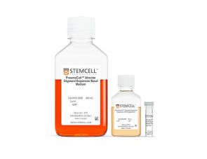
  • PneumaCult™-Ex Plus 基础培养基,490 mL
  • PneumaCult™-Ex Plus 50X 补充剂,10 mL
Interested in trying STEMCELL's products for respiratory research? Fill out this form to request information about introductory offers.
专为您的实验方案打造的产品
要查看实验方案所需的所有配套产品,请参阅《实验方案与技术文档》

总览

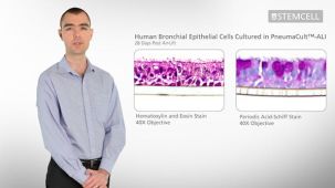
PneumaCult™-Ex Plus 培养基是一款无血清和无BPE的细胞培养基,与其他市售扩增培养基相比,它支持人原代呼吸道上皮细胞和鼻上皮细胞在每个代次均有更多的扩增。该培养基还支持至少两次额外的细胞扩增传代,并更好地维持细胞分化潜能,即在气液界面(ALI)培养条件下,使用PneumaCult™-ALI培养基(产品号 #05001)可形成假复层粘液纤毛上皮,或使用PneumaCult™-ALI-S培养基(产品号 #05050)可形成立方上皮的能力。

PneumaCult™-Ex Plus 培养基与PneumaCult™-ALI 培养基或PneumaCult™-ALI-S培养基共同构成了一个一体化的无 BPE 的体外人呼吸道培养模型。该培养模型效果稳定且成分明确,是呼吸系统基础研究、毒性研究和药物开发的宝贵工具。

通过我们的免费点播肺部课程学习如何在气液界面培养人呼吸道上皮细胞,或浏览关于使用PneumaCult™进行气液界面培养的常见问题解答(FAQ)。


分类
专用培养基
 
细胞类型
气道细胞
 
种属

 
应用
细胞培养,扩增,培养
 
品牌
PneumaCult
 
研究领域
上皮细胞研究
 
制剂类别
无血清
 

实验数据

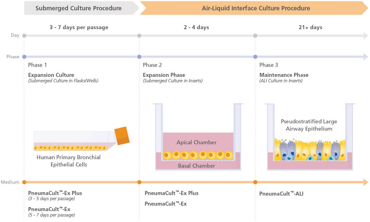
Figure 1. Overview of the PneumaCult™ culture system

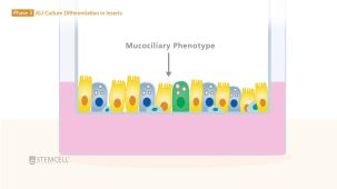
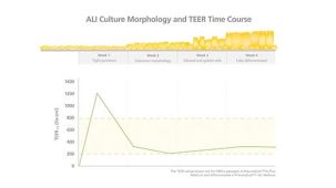
Expansion of human bronchial epithelial cells (HBECs) in submerged culture is performed with PneumaCult™-Ex Plus or PneumaCult™-Ex. During the early “Expansion Phase” of the air-liquid interface (ALI) culture procedure, PneumaCult™-Ex Plus or PneumaCult™-Ex is applied to the apical and basal chambers. Upon reaching confluence, the culture is air-lifted by removing the culture medium from both chambers, and adding PneumaCult™-ALI to the basal chamber only. Differentiation into a pseudostratified mucociliary epithelium is obtained following 21-28 days of incubation and can be maintained for more than one year.

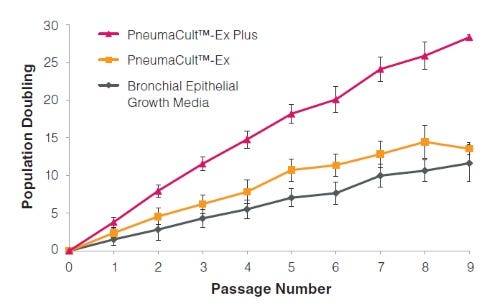
Figure 2. HBECs cultured in PneumaCult™-Ex Plus have a faster expansion rate compared to those cultured in PneumaCult™-Ex and Bronchial Epithelial Growth Media

Commercially available, cryopreserved P1 HBECs were seeded into PneumaCult™-Ex Plus, PneumaCult™-Ex, or Bronchial Epithelial Growth Media. Cells cultured in PneumaCult™-Ex Plus have a significantly higher proliferation rate over 9 passages compared to those maintained in either control medium (n=6).

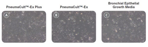
Figure 3. Representative morphology of HBECs

Representative live culture images for P4 HBECs cultured in PneumaCult™-Ex Plus, PneumaCult™-Ex, or Bronchial Epithelial Growth Media. Cells cultured in PneumaCult™-Ex Plus (A) are smaller and more tightly packed than those cultured in PneumaCult™-Ex (B) or Bronchial Epithelial Growth Media (C). All images were taken using a 10X objective.

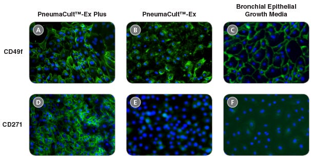
Figure 4. HBECs cultured in PneumaCult™-Ex Plus maintain widespread expression of the basal cell markers CD49f and CD271

Immunocytochemistry detection of basal cell markers - CD49f (A, B, and C) and CD271 (D, E, and F) - for P4 HBECs cultured in PneumaCult™-Ex Plus (A and D), PneumaCult™-Ex (B and E), and Bronchial Epithelial Growth Media (C and F). All images were taken using a 10X objective.

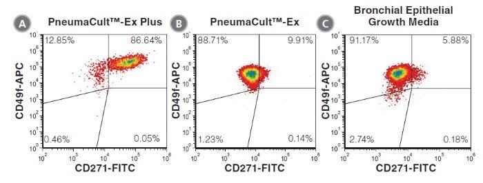
Figure 5. HBECs cultured in PneumaCult™-Ex Plus have a higher proportion of CD271+CD49f+ cells

P4 HBECs cultured in PneumaCult™-Ex Plus (A), PneumaCult™-Ex (B), and Bronchial Epithelial Growth Media (C) were characterized by flow cytometry to detect expression of the basal cell markers CD49f and CD271. HBECs cultured in PneumaCult™-Ex Plus (A) have a higher proportion of cells coexpressing CD49f and CD271, compared to those cultured in PneumaCult™-Ex (B) and Bronchial Epithelial Growth Media (C).

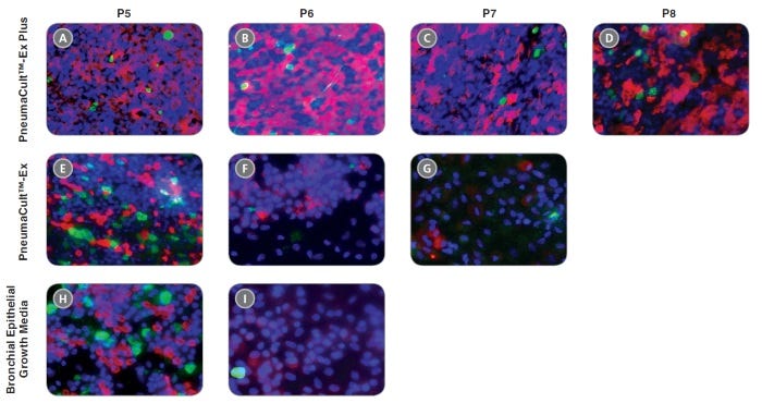
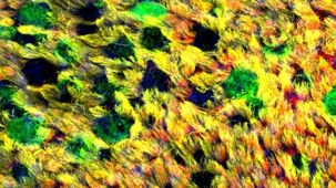
Figure 6. HBECs cultured in PneumaCult™-Ex Plus differentiate into a pseudostratified mucociliary epithelium at later passages with the use of PneumaCult™-ALI

P4 HBECs were seeded and passaged using PneumaCult™-Ex Plus, PneumaCult™-Ex, or Bronchial Epithelial Growth Media, followed by ALI differentiation at each passage (P5-8) with the use of PneumaCult™-ALI. The ALI cultures at 28 days post air-lift were fixed and stained with antibodies for cilia marker AC-tubulin (red) and the goblet cell marker Muc5AC (green). The nuclei are counterstained with DAPI (blue). All images were taken using a 20X objective.

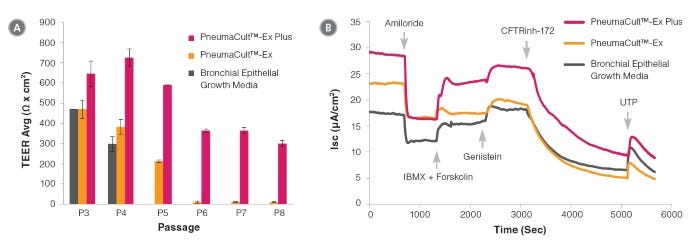
Figure 7. Electrophysiological characterization of differentiated HBECs (P4) that were expanded in PneumaCult™-Ex Plus, PneumaCult™-Ex, and Bronchial Epithelial Growth Media

Transepithelial electrical resistance (TEER) (A) and representative characterization of the ion channel activities (B) for ALI cultures at 28 days post air-lift using HBECs expanded in PneumaCult™-Ex Plus, PneumaCult™-Ex, or Bronchial Epithelial Growth Media. Amiloride: ENaC inhibitor. IBMX and Forskolin: CFTR activators. Genistein: CFTR potentiator. CFTRinh-172: CFTR inhibitor. UTP: Calciumactivated Chloride channels (CaCCs) activator. All ALI differentiation cultures were performed using PneumaCult™-ALI.

产品说明书及文档

请在《产品说明书》中查找相关支持信息和使用说明,或浏览下方更多实验方案。

Document Type
Product Name
Catalog #
Lot #
Language
Catalog #
05040
Lot #
All
Language
English
Document Type
Safety Data Sheet 1
Catalog #
05040
Lot #
All
Language
English
Document Type
Safety Data Sheet 2
Catalog #
05040
Lot #
All
Language
English

应用领域

本产品专为以下研究领域设计,适用于工作流程中的高亮阶段。探索这些工作流程,了解更多我们为各研究领域提供的其他配套产品。

相关材料与文献

技术资料 (18)

文献 (41)

Nasal polyp fibroblasts modulate epithelial characteristics via Wnt signaling. A. Dobzanski et al. International forum of allergy {\&} rhinology 2018 dec

Abstract

BACKGROUND While essential to the normal differentiation of ciliated airway epithelial cells,upregulated Wnt signaling in chronic rhinosinusitis with nasal polyps (CRSwNP) has been proposed to result in abnormal epithelial morphology and dysfunctional mucociliary clearance. The mechanism of epithelial Wnt signaling dysregulation in CRSwNP is unknown,and importantly cellular sources of Wnt ligands in CRSwNP have not yet been investigated. METHODS Human sinonasal epithelial cells (hSNECs) and human sinonasal fibroblasts (hSNFs) were collected from 34 human subjects (25 control and 9 CRSwNP) and differentiated as primary air-liquid interface (ALI) and organoid co-cultures. hSNECs were isolated to the apical compartment of the transwell and hSNFs were isolated to the basolateral compartment. After 21 days of ALI culture,ciliary expression and sinonasal epithelial morphology were examined by immunohistochemistry (IHC) and quantitative real-time polymerase chain reaction (qRT-PCR). An organoid model was used to evaluate proliferation of basal cells in presence of hSNFs. RESULTS Epithelial cells co-cultured with CRSwNP-hSNFs revealed significantly decreased ciliated cells,altered epithelial cell morphology,and increased colony forming efficiency compared to epithelial cells co-cultured with control-hSNFs. CRSwNP-hSNFs showed significantly higher messenger RNA (mRNA) expression of canonical WNT3A. A Wnt agonist,CHIR99021,replicated CRSwNP-hSNF co-cultures,and treatment with the Wnt inhibitor IWP2 prevented abnormal morphologies. CONCLUSION These results suggest that abnormal interactions between epithelial cells and fibroblasts may contribute to CRSwNP pathogenesis and supports the concept that dysregulated Wnt signaling contributes impairment to epithelial function in CRSwNP.
Optimization of Normal Human Bronchial Epithelial (NHBE) Cell 3D Cultures for in vitro Lung Model Studies. R. E. Rayner et al. Scientific reports 2019

Abstract

Robust in vitro lung models are required for risk assessment to measure key events leading to respiratory diseases. Primary normal human bronchial epithelial cells (NHBE) represent a good lung model but obtaining well-differentiated 3D cultures can be challenging. Here,we evaluated the ability to expand primary NHBE cells in different culture conditions while maintaining their 3D culture characteristics such as ciliated and goblet cells,and ion channel function. Differentiated cultures were optimally obtained with PneumaCult-Ex Plus (expansion medium)/PneumaCult-ALI (differentiation medium). Primary cells passaged up to four times maintained airway epithelial characteristics as evidenced by ciliated pseudostratified columnar epithelium with goblet cells,trans-epithelial electrical resistance (TEER) ({\textgreater}400 Ohms.cm2),and cystic fibrosis transmembrane conductance regulator-mediated short-circuit currents ({\textgreater}3 µA/cm2). No change in ciliary beat frequency (CBF) or airway surface liquid (ASL) meniscus length was observed up to passage six. For the first time,this study demonstrates that CFTR ion channel function and normal epithelial phenotypic characteristics are maintained in passaged primary NHBE cells. Furthermore,this study highlights the criticality of evaluating expansion and differentiation conditions for achieving optimal phenotypic and functional endpoints (CBF,ASL,ion channel function,presence of differentiated cells,TEER) when developing in vitro lung models.
Neutrophils from severe asthmatic patients induce epithelial to mesenchymal transition in healthy bronchial epithelial cells. A. Haddad et al. Respiratory research 2019 oct

Abstract

BACKGROUND Asthma is a heterogenous disease characterized by chronic inflammation and airway remodeling. An increase in the severity of airway remodeling is associated with a more severe form of asthma. There is increasing interest in the epithelial to mesenchymal transition process and mechanisms involved in the differentiation and repair of the airway epithelium,especially as they apply to severe asthma. Growing evidence suggests that Epithelial-Mesenchymal transition (EMT) could contribute to airway remodeling and fibrosis in asthma. Severe asthmatic patients with remodeled airways have a neutrophil driven inflammation. Neutrophils are an important source of TGF-$\beta$1,which plays a role in recruitment and activation of inflammatory cells,extracellular matrix (ECM) production and fibrosis development,and is a potent inducer of EMT. OBJECTIVE As there is little data examining the contribution of neutrophils and/or their mediators to the induction of EMT in airway epithelial cells,the objective of this study was to better understand the potential role of neutrophils in severe asthma in regards to EMT. METHODS We used an in vitro system to investigate the neutrophil-epithelial cell interaction. We obtained peripheral blood neutrophils from severe asthmatic patients and control subjects and examined for their ability to induce EMT in primary airway epithelial cells. RESULTS Our data indicate that neutrophils from severe asthmatic patients induce changes in morphology and EMT marker expression in bronchial epithelial cells consistent with the EMT process when co-cultured. TGF-$\beta$1 levels in the culture medium of severe asthmatic patients were increased compared to that from co-cultures of non-asthmatic neutrophils and epithelial cells. CONCLUSIONS AND CLINICAL RELEVANCE As an inducer of EMT and an important source of TGF-$\beta$1,neutrophils may play a significant role in the development of airway remodeling and fibrosis in severe asthmatic airways.

更多信息

更多信息
物种
配方 无血清

法律声明:

This product was developed under a license to intellectual property owned or controlled by Propagenix, Inc. This product is sold for research use only (which includes pre-clinical research) under a non-transferable, limited-use license. Purchase of this product does not include the right to sell, use or otherwise transfer this product for commercial purposes or clinical use. Purchasers wishing to use the product for purposes other than research use should contact Propagenix, Inc. (www.propagenix.com/about#contact-us).

质量保证:

产品仅供研究使用,不用于针对人或动物的诊断或治疗。 欲获悉更多关于STEMCELL的质控信息,请访问 STEMCELL.CN/COMPLIANCE.

Copyright © 2026 by STEMCELL Technologies. All rights reserved.

在线联系