若您需要咨询产品或有任何技术问题,请通过官方电话 400 885 9050 或邮箱 info.cn@stemcell.com 与我们联系。

PneumaCult™-ALI 培养基

用于在气液界面培养的人呼吸道上皮细胞的无血清和无BPE培养基
只有 %1
¥3,570.00

产品号 #(选择产品)

产品号 #05001_C

用于在气液界面培养的人呼吸道上皮细胞的无血清和无BPE培养基

产品优势

  • 用PneumaCult™-ALI培养的HBECs经过广泛的粘膜纤毛分化,形成与人呼吸道非常相似的假复层上皮结构
  • PneumaCult™-ALI不含血清和BPE,可最大限度地减少差异

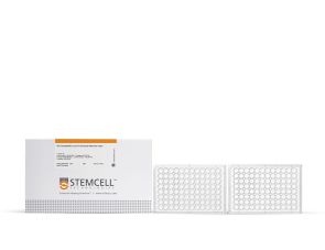
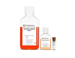
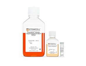
产品组分包括

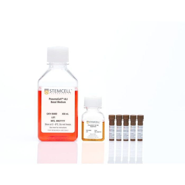
  • PneumaCult™-ALI 基础培养基,450 mL
  • PneumaCult™-ALI 10X 补充剂, 50 mL
  • PneumaCult™-ALI 维持补充剂 (100X), 5 x 1 mL
Interested in trying STEMCELL's products for respiratory research? Fill out this form to request information about introductory offers.
专为您的实验方案打造的产品
要查看实验方案所需的所有配套产品,请参阅《实验方案与技术文档》

总览

PneumaCult™-ALI 培养基(产品号#05001)是一款不含血清和BPE的培养基,用于在气液界面(ALI)培养人呼吸道上皮细胞。在PneumaCult™-ALI 培养基中培养的呼吸道上皮细胞经过广泛的粘膜纤毛分化形成假复层上皮,其形态和功能特征与体内的人呼吸道相似。PneumaCult™-ALI Medium也可提供套装,包括12毫米Transwell®插件(产品号#05021)或6.5毫米Transwell®插件(产品号#05022)。

PneumaCult™-ALI 培养基和PneumaCult™-NGEx培养基(产品号#100-1505)构成了一个完整、无BPE的人源呼吸道上皮体外建模系统,也与原代人鼻上皮细胞兼容。这一稳定、定义明确的培养系统是开展基础呼吸研究、毒理学研究和药物开发的宝贵工具。

欢迎观看我们的肺部研究课程,了解如何在 ALI 条件下培养人呼吸道上皮细胞,或浏览我们的常见问题解答(FAQs)关于使用PneumaCult™的ALI培养工作流程。


分类
专用培养基
 
细胞类型
气道细胞
 
种属

 
应用
细胞培养,分化,培养,类器官培养
 
品牌
PneumaCult
 
研究领域
疾病建模,药物发现和毒理检测,上皮细胞研究
 
制剂类别
无血清
 

实验数据

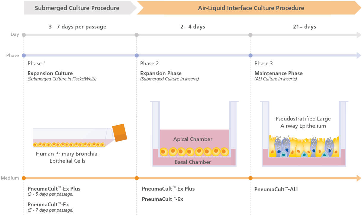
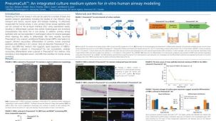
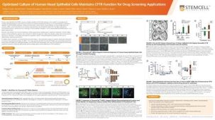
Figure 1. Overview of the PneumaCult™ Culture System

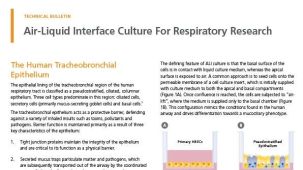
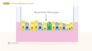
Expansion of human bronchial epithelial cells (HBECs) in submerged culture is performed with PneumaCult™-Ex Plus or PneumaCult™-Ex. During the early Expansion Phase of the ALI culture procedure, PneumaCult™-Ex Plus or PneumaCult™-Ex is applied to the apical and basal chambers. Upon reaching confluence, the culture is air-lifted by removing the culture medium from both chambers, and adding PneumaCult™-ALI to the basal chamber only. Differentiation into a pseudostratified mucociliary epithelium is obtained following 21-28 days of incubation and can be maintained for more than one year.

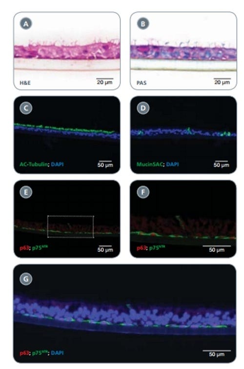
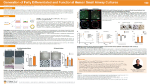
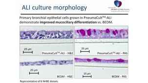
Figure 2. HBECs Cultured in PneumaCult™-Ex Successfully Differentiate into a Pseudostratified Mucociliary Epithelium with PneumaCult™-ALI

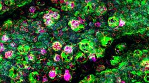
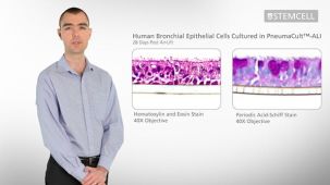
Early-passage (P1-3) HBECs cultured in PneumaCult™-Ex successfully differentiate when cultured at air-liquid interface with PneumaCult™-ALI for 28 days. H&E staining revealed the pseudostratifi ed structure of the epithelium with cilia present at the apical surface (A). Periodic acid-Schiff staining demonstrated the presence of goblet cells (B). The presence of ciliated and goblet cells was also demonstrated by immunofl uorescence staining of cilia marker acetylated (AC)-Tubulin (green; C) and the goblet cell marker Mucin5AC (green; D). Appropriate positioning of basal cells along the transwell insert was visualized by immunofl uorescence staining using the basal cell markers p75NTR (green) and p63 (red; E,F). A representative merged image indicates the apical cells, detected by DAPI alone, positioned along the epithelium and in close contact with the basal cells (detected by DAPI, p63 and p75NTR co-labeling) located along the insert (G).

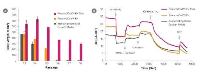
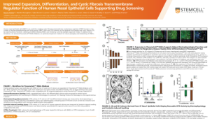
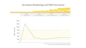
Figure 3. Electrophysiological characterization of differentiated HBECs (P4) that were expanded in PneumaCult™-Ex Plus, PneumaCult™-Ex, and Bronchial Epithelial Growth Media

TEER (A) and representative characterization of the ion channel activities (B) for ALI cultures at 28 days post air-lift using HBECs expanded in PneumaCult™-Ex Plus, PneumaCult™-Ex, or Bronchial Epithelial Growth Media. Amiloride: ENaC inhibitor. IBMX and Forskolin: CFTR activators. Genistein: CFTR potentiator. CFTRinh-172: CFTR inhibitor. UTP: Calciumactivated Chloride channels (CaCCs) activator. All ALI differentiation cultures were performed using PneumaCult™-ALI.

产品说明书及文档

请在《产品说明书》中查找相关支持信息和使用说明,或浏览下方更多实验方案。

Document Type
Product Name
Catalog #
Lot #
Language
Catalog #
05001
Lot #
All
Language
English
Catalog #
05021
Lot #
All
Language
English
Catalog #
05022
Lot #
All
Language
English
Document Type
Safety Data Sheet 1
Catalog #
05001
Lot #
All
Language
English
Document Type
Safety Data Sheet 2
Catalog #
05001
Lot #
All
Language
English
Document Type
Safety Data Sheet 3
Catalog #
05001
Lot #
All
Language
English
Document Type
Safety Data Sheet 1
Catalog #
05021
Lot #
All
Language
English
Document Type
Safety Data Sheet 2
Catalog #
05021
Lot #
All
Language
English
Document Type
Safety Data Sheet 3
Catalog #
05021
Lot #
All
Language
English
Document Type
Safety Data Sheet 1
Catalog #
05022
Lot #
All
Language
English
Document Type
Safety Data Sheet 2
Catalog #
05022
Lot #
All
Language
English
Document Type
Safety Data Sheet 3
Catalog #
05022
Lot #
All
Language
English

应用领域

本产品专为以下研究领域设计,适用于工作流程中的高亮阶段。探索这些工作流程,了解更多我们为各研究领域提供的其他配套产品。

相关材料与文献

技术资料 (27)

产品手册

文献 (124)

Culturing of human nasal epithelial cells at the air liquid interface. Mü et al. Journal of visualized experiments : JoVE 2013 JAN

Abstract

In vitro models using human primary epithelial cells are essential in understanding key functions of the respiratory epithelium in the context of microbial infections or inhaled agents. Direct comparisons of cells obtained from diseased populations allow us to characterize different phenotypes and dissect the underlying mechanisms mediating changes in epithelial cell function. Culturing epithelial cells from the human tracheobronchial region has been well documented,but is limited by the availability of human lung tissue or invasiveness associated with obtaining the bronchial brushes biopsies. Nasal epithelial cells are obtained through much less invasive superficial nasal scrape biopsies and subjects can be biopsied multiple times with no significant side effects. Additionally,the nose is the entry point to the respiratory system and therefore one of the first sites to be exposed to any kind of air-borne stressor,such as microbial agents,pollutants,or allergens. Briefly,nasal epithelial cells obtained from human volunteers are expanded on coated tissue culture plates,and then transferred onto cell culture inserts. Upon reaching confluency,cells continue to be cultured at the air-liquid interface (ALI),for several weeks,which creates more physiologically relevant conditions. The ALI culture condition uses defined media leading to a differentiated epithelium that exhibits morphological and functional characteristics similar to the human nasal epithelium,with both ciliated and mucus producing cells. Tissue culture inserts with differentiated nasal epithelial cells can be manipulated in a variety of ways depending on the research questions (treatment with pharmacological agents,transduction with lentiviral vectors,exposure to gases,or infection with microbial agents) and analyzed for numerous different endpoints ranging from cellular and molecular pathways,functional changes,morphology,etc. In vitro models of differentiated human nasal epithelial cells will enable investigators to address novel and important research questions by using organotypic experimental models that largely mimic the nasal epithelium in vivo.
Dedifferentiation of committed epithelial cells into stem cells in vivo. Tata PR et al. Nature 2013 NOV

Abstract

Cellular plasticity contributes to the regenerative capacity of plants,invertebrates,teleost fishes and amphibians. In vertebrates,differentiated cells are known to revert into replicating progenitors,but these cells do not persist as stable stem cells. Here we present evidence that differentiated airway epithelial cells can revert into stable and functional stem cells in vivo. After the ablation of airway stem cells,we observed a surprising increase in the proliferation of committed secretory cells. Subsequent lineage tracing demonstrated that the luminal secretory cells had dedifferentiated into basal stem cells. Dedifferentiated cells were morphologically indistinguishable from stem cells and they functioned as well as their endogenous counterparts in repairing epithelial injury. Single secretory cells clonally dedifferentiated into multipotent stem cells when they were cultured ex vivo without basal stem cells. By contrast,direct contact with a single basal stem cell was sufficient to prevent secretory cell dedifferentiation. In analogy to classical descriptions of amphibian nuclear reprogramming,the propensity of committed cells to dedifferentiate is inversely correlated to their state of maturity. This capacity of committed cells to dedifferentiate into stem cells may have a more general role in the regeneration of many tissues and in multiple disease states,notably cancer.
Cigarette smoke-induced disruption of bronchial epithelial tight junctions is prevented by transforming growth factor-beta. Schamberger AC et al. American journal of respiratory cell and molecular biology 2014 JUN

Abstract

The airway epithelium constitutes an essential immunological and cytoprotective barrier to inhaled insults,such as cigarette smoke,environmental particles,or viruses. Although bronchial epithelial integrity is crucial for airway homeostasis,defective epithelial barrier function contributes to chronic obstructive pulmonary disease (COPD). Tight junctions at the apical side of epithelial cell-cell contacts determine epithelial permeability. Cigarette smoke exposure,the major risk factor for COPD,is suggested to impair tight junction integrity; however,detailed mechanisms thereof remain elusive. We investigated whether cigarette smoke extract (CSE) and transforming growth factor (TGF)-$$1 affected tight junction integrity. Exposure of human bronchial epithelial cells (16HBE14o(-)) and differentiated primary human bronchial epithelial cells (pHBECs) to CSE significantly disrupted tight junction integrity and barrier function. Specifically,CSE decreased transepithelial electrical resistance (TEER) and tight junction-associated protein levels. Zonula occludens (ZO)-1 and ZO-2 protein levels were significantly reduced and dislocated from the cell membrane,as observed by fractionation and immunofluorescence analysis. These findings were reproduced in isolated bronchi exposed to CSE ex vivo,as detected by real-time quantitative reverse-transcriptase PCR and immunohistochemistry. Combined treatment of 16HBE14o(-) cells or pHBECs with CSE and TGF-$$1 restored ZO-1 and ZO-2 levels. TGF-$$1 cotreatment restored membrane localization of ZO-1 and ZO-2 protein and prevented CSE-mediated TEER decrease. In conclusion,CSE led to the disruption of tight junctions of human bronchial epithelial cells,and TGF-$$1 counteracted this CSE-induced effect. Thus,TGF-$$1 may serve as a protective factor for bronchial epithelial cell homeostasis in diseases such as COPD.

更多信息

更多信息
物种
配方 无血清
质量保证:

产品仅供研究使用,不用于针对人或动物的诊断或治疗。 欲获悉更多关于STEMCELL的质控信息,请访问 STEMCELL.CN/COMPLIANCE.
Copyright © 2026 by STEMCELL Technologies. All rights reserved.

在线联系