若您需要咨询产品或有任何技术问题,请通过官方电话 400 885 9050 或邮箱 info.cn@stemcell.com 与我们联系。

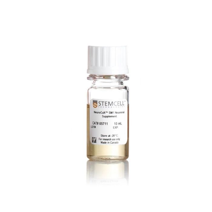
NeuroCult™ SM1 神经添加物

无血清神经添加物(50X)
只有 %1
¥1,384.00

产品号 #(选择产品)

产品号 #05711_C

无血清神经添加物(50X)

产品优势

  • 多功能细胞培养添加物
  • 优化的无血清配方
  • 原材料经过严格筛选,保证批次间的一致性
专为您的实验方案打造的产品
要查看实验方案所需的所有配套产品,请参阅《实验方案与技术文档》

总览

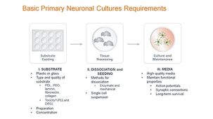
使用基于 Brewer's B27 的添加物,可为中枢神经系统 (CNS) 衍生细胞或人多能干细胞 (hPSC) 衍生细胞提供稳定的神经元培养体系,从而避免培养失败。NeuroCult™ SM1 (STEMCELL Modified-1) 基于已公开的配方 (Brewer et al. J Neurosci Res., 1993) 经过标准化,可更稳定地支持原代和人多能干细胞(hPSC)来源的神经元的存活和成熟。

这款无血清添加物可以与基础培养基和各种不同的诱导因子或细胞因子一起使用,以支持外胚层、中胚层和内胚层谱系的分化。NeuroCult™SM1也可用作血清替代补充剂,如神经毒性测定和钙成像。

为了您的方便使用,NeuroCult™SM1包含在多个BrainPhys™神经元培养基试剂盒中,用于原代和hPSC衍生的神经元(产品号 #05792,05793,05794和05795)。更多详情,请参阅下方BrainPhys™的性能数据。

包含
• 抗氧化剂
• 维生素A
• 胰岛素
• 其他成分
 
分类
添加剂
 
细胞类型
神经细胞,PSC衍生,神经元,多能干细胞
 
种属
人,小鼠,大鼠
 
应用
细胞培养,分化,培养
 
品牌
NeuroCult
 
研究领域
药物发现和毒性检测,神经科学,干细胞生物学
 
制剂类别
无血清
 

实验数据

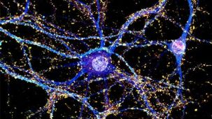
Morphology of Neurons in Representative NeuroCult™ SM1 Cultures at 7 and 21 Days in Vitro

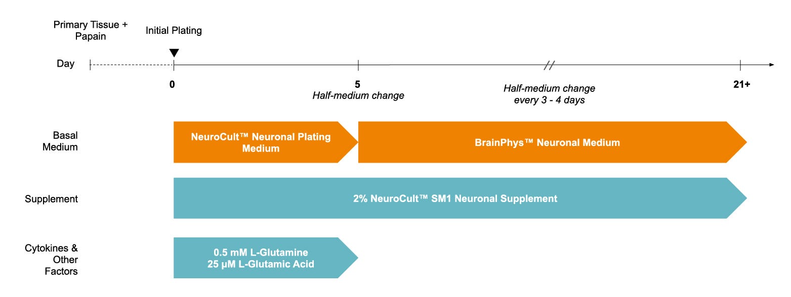
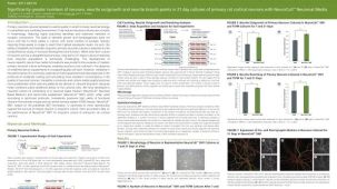
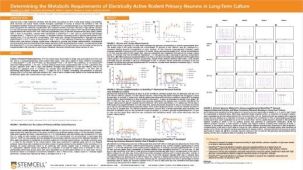
Figure 1. Protocol for Plating and Culturing Primary Neurons with the SM1 Culture System

Primary rodent tissue dissociated in papain was plated in NeuroCult™ Neuronal Plating Medium, supplemented with NeuroCult™ SM1 Neuronal Supplement, L-Glutamine, and L-Glutamic Acid. On day 5, primary neurons were transitioned to BrainPhys™ Neuronal Medium, supplemented with NeuroCult™ SM1 Neuronal Supplement, by performing half-medium changes every 3 - 4 days.

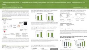
Number of Neurons in NeuroCult™ SM1 and TSFM Cultures After 7 and 21 Days in Vitro

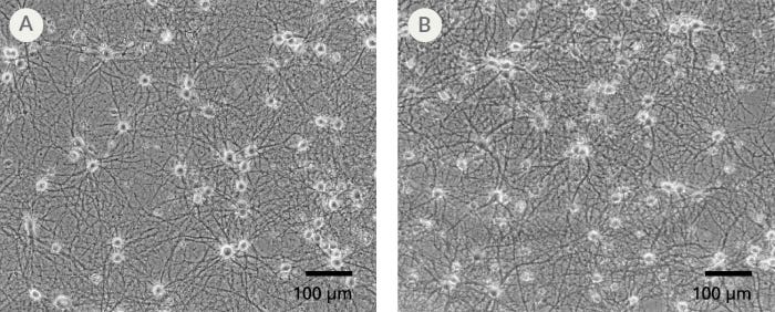
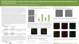
Figure 2. The SM1 Culture System Supports Long-Term Culture of Rodent Neurons

Primary E18 rat cortical neurons were cultured in the SM1 Culture System. A large number of viable neurons are visible after (A) 21 and (B) 35 days, as demonstrated by their bright neuronal cell bodies, and extensive neurite outgrowth and branching. Neurons are evenly distributed over the culture surface with minimal cell clumping.

Neurite Outgrowth of Primary Neurons Cultured in NeuroCult™ SM1 and TSFM for 7 and 21 Days

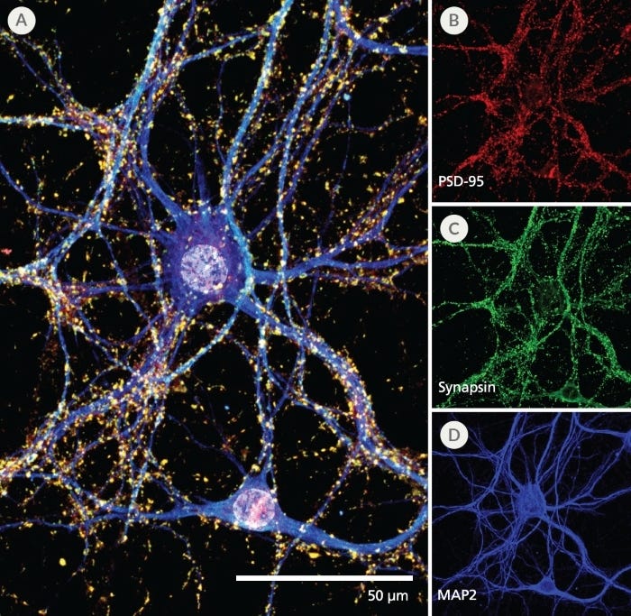
Figure 3. Pre- and Post-Synaptic Markers are Expressed in Rodent Neurons Cultured in the SM1 Culture System

Primary E18 rat cortical neurons were cultured in the SM1 Culture System. At 21 DIV, neurons are phenotypically mature, as indicated by the presence of an extensive dendritic arbor, and appropriate expression and localization of pre-synaptic synapsin (A,C; green) and post-synaptic PSD-95 (A,B; red) markers. Synapsin is concentrated in discrete puncta distributed along the somata and dendritic processes, as defined by the dendritic marker MAP2 (A,D; blue).

MEA data showing mean firing rate of rodent primary neurons cultured in BrainPhys™ and other commercial media

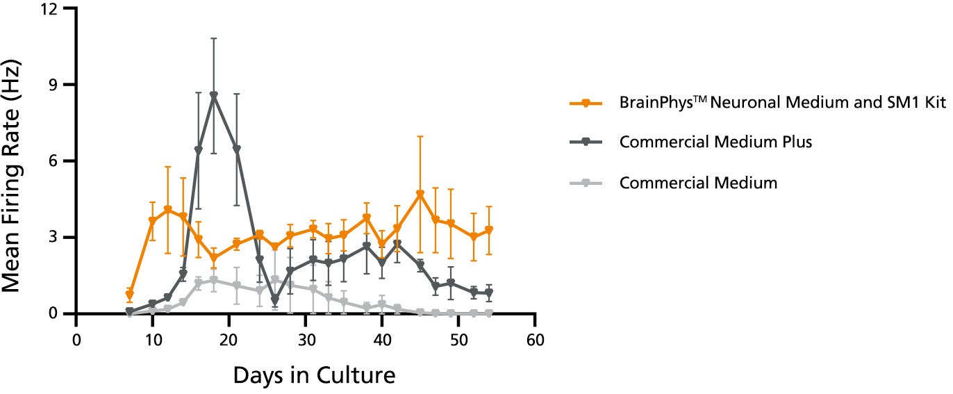
Figure 4. Glucose Supplementation in BrainPhys™ Maintains Neuronal Activity Over 8 Weeks in Culture

Primary E18 rat cortical neurons were cultured with BrainPhys™ and SM1 or other commercially available culture systems for 8 weeks. Neuronal activity can be detected at Day 9 with BrainPhys™, whereas activity is not detected until Day 14 in cultures maintained in either of the Commercial Media with Commercial Supplements. For Commercial Medium and Supplement-cultured neurons, mean firing rate remains low throughout culture. In contrast, a “peak-drop” activity pattern is observed in the Commercial Medium Plus condition, where mean firing rate increases rapidly within 2 days, followed by a drop in activity in the next 2 - 4 days. BrainPhys™and SM1 Kit with 15 mM glucose maintains the highest level of activity throughout the 8-week culture period.

Raster plots showing activity of neurons cultured in BrainPhys™ and other commercial media

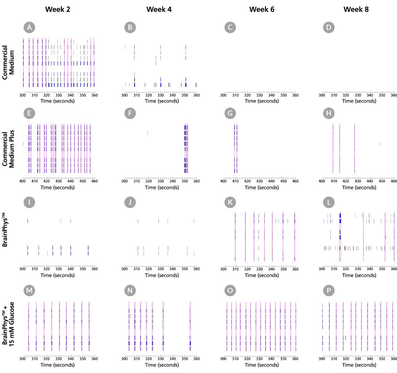
Figure 5. BrainPhys™ Supports Improved Neuronal Activity and More Consistent Network Bursting in Long-Term Culture

Raster plots from MEA recordings show the firing patterns of neurons across 8 electrodes at Weeks 2, 4, 6 and 8. Neurons were either cultured with a Commercial Medium with Supplements, Commercial Medium Plus with Supplements, BrainPhys™ and SM1, or BrainPhys™ and SM1 with 15 mM glucose. Detected spikes (black lines), single channel bursts (blue lines; a collection of at least 5 spikes, each separated by an ISI of no more than 100 ms), and network bursts (magenta boxes; a collection of at least 50 spikes from a minimum of 35% of participating electrodes across each well, each separated by an ISI of no more than 100 ms) were recorded for each medium. (A-D) Neurons cultured with Commercial Medium exhibited network bursting in Week 2 but no spiking activity was detected in subsequent timepoints. (E-H) In Commercial Medium Plus-cultured neurons, a high number of spikes and regular network bursting were detected at Week 2. A decreased number of spikes and inconsistent network bursting were observed in later time points, corresponding to the drop in MFR seen in Figure 4. (I-L) Without glucose, individual spiking was observed at Weeks 2 and 4 with BrainPhys™ and SM1 but network bursting was not detected until Weeks 6 and 8. (M-T) In contrast, neurons cultured with BrainPhys™ and SM1 with 15 mM glucose demonstrated strong spiking activity and consistent network bursting at all timepoints. MEA = microelectrode array; ISI = inter-spike interval; MFR = mean firing rate

产品说明书及文档

请在《产品说明书》中查找相关支持信息和使用说明,或浏览下方更多实验方案。

Document Type
Product Name
Catalog #
Lot #
Language
Catalog #
100-1281
Lot #
All
Language
English
Catalog #
05711
Lot #
All
Language
English
Document Type
Safety Data Sheet
Catalog #
100-1281
Lot #
All
Language
English
Document Type
Safety Data Sheet
Catalog #
05711
Lot #
All
Language
English

相关材料与文献

技术资料 (15)

文献 (59)

Inhibitory synapse dynamics: coordinated presynaptic and postsynaptic mobility and the major contribution of recycled vesicles to new synapse formation. Dobie FA and Craig AM The Journal of neuroscience : the official journal of the Society for Neuroscience 2011 JUL

Abstract

Dynamics of GABAergic synaptic components have been studied previously over milliseconds to minutes,revealing mobility of postsynaptic scaffolds and receptors. Here we image inhibitory synapses containing fluorescently tagged postsynaptic scaffold Gephyrin,together with presynaptic vesicular GABA transporter (VGAT) or postsynaptic GABA(A) receptor γ2 subunit (GABA(A)Rγ2),over seconds to days in cultured rat hippocampal neurons,revealing modes of inhibitory synapse formation and remodeling. Entire synapses were mobile,translocating rapidly within a confined region and exhibiting greater nonstochastic motion over multihour periods. Presynaptic and postsynaptic components moved in unison,maintaining close apposition while translocating distances of several micrometers. An observed flux in the density of synaptic puncta partially resulted from the apparent merging and splitting of preexisting clusters. De novo formation of inhibitory synapses was observed,marked by the appearance of stably apposed Gephyrin and VGAT clusters at sites previously lacking either component. Coclustering of GABA(A)Rγ2 supports the identification of such new clusters as synapses. Nascent synapse formation occurred by gradual accumulation of components over several hours,with VGAT clustering preceding that of Gephyrin and GABA(A)Rγ2. Comparing VGAT labeling by active uptake of a luminal domain antibody with post hoc immunocytochemistry indicated that recycling vesicles from preexisting boutons significantly contribute to vesicle pools at the majority of new inhibitory synapses. Although new synapses formed primarily on dendrite shafts,some also formed on dendritic protrusions,without apparent interconversion. Altogether,the long-term imaging of GABAergic presynaptic and postsynaptic components reveals complex dynamics and perpetual remodeling with implications for mechanisms of assembly and synaptic integration.
NMDA receptors mediate synaptic competition in culture. She K and Craig AM PloS one 2011 JAN

Abstract

BACKGROUND: Activity through NMDA type glutamate receptors sculpts connectivity in the developing nervous system. This topic is typically studied in the visual system in vivo,where activity of inputs can be differentially regulated,but in which individual synapses are difficult to visualize and mechanisms governing synaptic competition can be difficult to ascertain. Here,we develop a model of NMDA-receptor dependent synaptic competition in dissociated cultured hippocampal neurons. METHODOLOGY/PRINCIPAL FINDINGS: GluN1 -/- (KO) mouse hippocampal neurons lacking the essential NMDA receptor subunit were cultured alone or cultured in defined ratios with wild type (WT) neurons. The absence of functional NMDA receptors did not alter neuron survival. Synapse development was assessed by immunofluorescence for postsynaptic PSD-95 family scaffold and apposed presynaptic vesicular glutamate transporter VGlut1. Synapse density was specifically enhanced onto minority wild type neurons co-cultured with a majority of GluN1 -/- neighbour neurons,both relative to the GluN1 -/- neighbours and relative to sister pure wild type cultures. This form of synaptic competition was dependent on NMDA receptor activity and not conferred by the mere physical presence of GluN1. In contrast to these results in 10% WT and 90% KO co-cultures,synapse density did not differ by genotype in 50% WT and 50% KO co-cultures or in 90% WT and 10% KO co-cultures. CONCLUSIONS/SIGNIFICANCE: The enhanced synaptic density onto NMDA receptor-competent neurons in minority coculture with GluN1 -/- neurons represents a cell culture paradigm for studying synaptic competition. Mechanisms involved may include a retrograde 'reward' signal generated by WT neurons,although in this paradigm there was no 'punishment' signal against GluN1 -/- neurons. Cell culture assays involving such defined circuits may help uncover the rules and mechanisms of activity-dependent synaptic competition in the developing nervous system.
The ADNP derived peptide, NAP modulates the tubulin pool: implication for neurotrophic and neuroprotective activities. Oz S et al. PloS one 2012 JAN

Abstract

Microtubules (MTs),key cytoskeletal elements in living cells,are critical for axonal transport,synaptic transmission,and maintenance of neuronal morphology. NAP (NAPVSIPQ) is a neuroprotective peptide derived from the essential activity-dependent neuroprotective protein (ADNP). In Alzheimer's disease models,NAP protects against tauopathy and cognitive decline. Here,we show that NAP treatment significantly affected the alpha tubulin tyrosination cycle in the neuronal differentiation model,rat pheochromocytoma (PC12) and in rat cortical astrocytes. The effect on tubulin tyrosination/detyrosination was coupled to increased MT network area (measured in PC12 cells),which is directly related to neurite outgrowth. Tubulin beta3,a marker for neurite outgrowth/neuronal differentiation significantly increased after NAP treatment. In rat cortical neurons,NAP doubled the area of dynamic MT invasion (Tyr-tubulin) into the neuronal growth cone periphery. NAP was previously shown to protect against zinc-induced MT/neurite destruction and neuronal death,here,in PC12 cells,NAP treatment reversed zinc-decreased tau-tubulin-MT interaction and protected against death. NAP effects on the MT pool,coupled with increased tau engagement on compromised MTs imply an important role in neuronal plasticity,protecting against free tau accumulation leading to tauopathy. With tauopathy representing a major pathological hallmark in Alzheimer's disease and related disorders,the current findings provide a mechanistic basis for further development. NAP (davunetide) is in phase 2/3 clinical trial in progressive supranuclear palsy,a disease presenting MT deficiency and tau pathology.

更多信息

更多信息
物种 人, 大鼠, 小鼠
Contains • Antioxidants • Vitamin A • Insulin • Other ingredients
配方 无血清
质量保证:

产品仅供研究使用,不用于针对人或动物的诊断或治疗。 欲获悉更多关于STEMCELL的质控信息,请访问 STEMCELL.CN/COMPLIANCE. Safety Statement: CA WARNING: This product can expose you to Progesterone which is known to the State of California to cause cancer. For more information go to www.P65Warnings.ca.gov
Copyright © 2026 by STEMCELL Technologies. All rights reserved.

在线联系