若您需要咨询产品或有任何技术问题,请通过官方电话 400 885 9050 或邮箱 info.cn@stemcell.com 与我们联系。

MesenCult™ 成骨刺激试剂盒(小鼠)

小鼠间充质干细胞和胚胎成纤维细胞分化为成骨细胞的完全培养基
只有 %1
¥5,652.00

产品号 #(选择产品)

产品号 #05504_C

小鼠间充质干细胞和胚胎成纤维细胞分化为成骨细胞的完全培养基

产品优势

  • 本试剂盒可诱导扩增过的小鼠间充质干祖细胞和胚胎成纤维细胞发生强力的成骨分化作用,这体现在参与骨分化和成熟的关键成骨转录基因的高表达以及茜素红染色上。参见 “数据和出版物 ”产品页面分栏。

产品组分包括

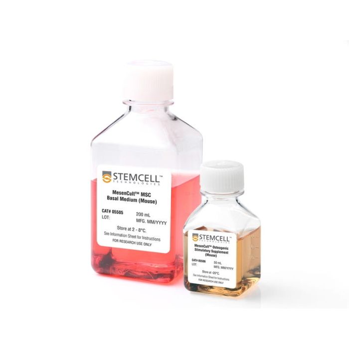
  • MesenCult™ MSC 基础培养基(小鼠),200 mL
  • MesenCult™ MSC成骨刺激补充剂(小鼠),50 mL
专为您的实验方案打造的产品
要查看实验方案所需的所有配套产品,请参阅《实验方案与技术文档》

总览

MesenCult™成骨刺激试剂盒(小鼠)是专为来自致密骨和骨髓的小鼠间充质干祖细胞(MSCs)以及小鼠胚胎成纤维细胞(MEFs)分化为成骨细胞而研制。本试剂盒可诱导小鼠MSCs和 MEFs 强力成骨,这一点可通过对参与骨分化和成熟的关键转录基因进行 qPCR 分析和茜素红染色得到证明。本试剂盒推荐用于鉴定MSCs和 MEFs 以及研究骨发育。

分类
专用培养基
 
细胞类型
间充质干/祖细胞,鼠胚胎成纤维细胞,成骨细胞
 
种属
小鼠
 
应用
细胞培养,分化
 
品牌
MesenCult
 
研究领域
药物发现和毒性检测,干细胞生物学
 

实验数据

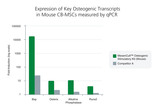
1 MOUSE COMPACT BONE-DERIVED MSC (CB-MSC) DATA<br>Compact bone-derived MSCs exhibited superior expression of key osteogenic transcripts involved in bone differentiation and maturation following treatment with MesenCult™ Osteogenic Stimulatory Kit (Mouse)*

Figure 1. Differentiation of mouse compact bone-derived mesenchymal stem cells (CB-MSCs) with the MesenCult™ Osteogenic Stimulatory Kit (Mouse)

Compact bone-derived MSCs exhibited superior expression of key osteogenic transcripts involved in bone differentiation and maturation following treatment with MesenCult™ Osteogenic Stimulatory Kit (Mouse)*

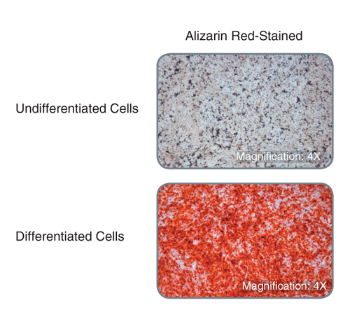
Compact bone-derived MSCs display robust bone matrix deposition following treatment with MesenCult™ Osteogenic Stimulatory Kit (Mouse) as demonstrated by Alizarin Red-staining

Figure 2. Compact bone-derived MSCs display robust bone matrix deposition following treatment with MesenCult™ Osteogenic Stimulatory Kit (Mouse) as demonstrated by Alizarin Red-staining

Ca2+ deposits are detected with Alizarin Red.
For both the qPCR analysis and Alizarin-Red staining assay, the mouse compact bone-derived MSCs were cultured in vitro for three passages, purified with EasySep™ Mesenchymal Stem/Progenitor Cell Enrichment Kit (Mouse, Catalog #19771), and then cultured for another passage, prior to differentiation with the MesenCult™ Osteogenic Stimulatory Kit (Mouse).

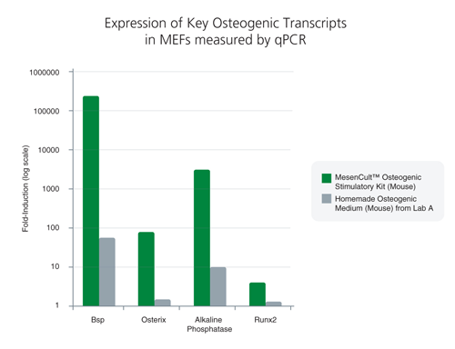
2 MOUSE EMBRYONIC FIBROBLASTS (MEF) DATA<br>MEFs exhibited superior expression of key osteogenic transcripts involved in bone differentiation and maturation following treatment with MesenCult™ Osteogenic Stimulatory Kit (Mouse)*

Figure 3.Differentiation of mouse embryonic fibroblasts (MEFs) with MesenCult™ Osteogenic Stiumulatory Kit (Mouse)

MEFs exhibited superior expression of key osteogenic transcripts involved in bone differentiation and maturation following treatment with MesenCult™ Osteogenic Stimulatory Kit (Mouse)*

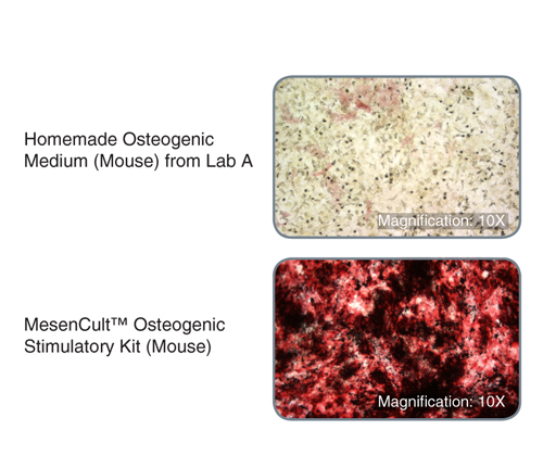
MEFs treated with MesenCult™ Osteogenic Stimulatory Kit (Mouse) displayed substantially increased alkaline phosphatase activity and bone matrix deposition*

Figure 4. MEFs treated with MesenCult™ Osteogenic Stimulatory Kit (Mouse) displayed substantially increased alkaline phosphatase activity and bone matrix deposition*

Alkaline phosphatase activity is shown by Naphthol AS-MX Phosphate/Fast Red Violet LB Salt staining (red areas) while bone matrix deposition is shown by von Kossa staining (black areas).
*Versus a homemade osteogenic medium (mouse) from Lab A.
For the qPCR analysis and staining assays, MEFs were cultured in vitro for 20 days using MesenCult™ Osteogenic Stimulatory Kit (Mouse), homemade, or MesenCult™ Proliferation Kit (Mouse, Catalog #05511) as the negative control.

产品说明书及文档

请在《产品说明书》中查找相关支持信息和使用说明,或浏览下方更多实验方案。

Document Type
Product Name
Catalog #
Lot #
Language
Catalog #
05504
Lot #
All
Language
English
Document Type
Safety Data Sheet 1
Catalog #
05504
Lot #
All
Language
English
Document Type
Safety Data Sheet 2
Catalog #
05504
Lot #
All
Language
English

应用领域

本产品专为以下研究领域设计,适用于工作流程中的高亮阶段。探索这些工作流程,了解更多我们为各研究领域提供的其他配套产品。

相关材料与文献

技术资料 (5)

文献 (6)

Hookworm Secreted Extracellular Vesicles Interact With Host Cells and Prevent Inducible Colitis in Mice. R. M. Eichenberger et al. Frontiers in immunology 2018

Abstract

Gastrointestinal (GI) parasites,hookworms in particular,have evolved to cause minimal harm to their hosts,allowing them to establish chronic infections. This is mediated by creating an immunoregulatory environment. Indeed,hookworms are such potent suppressors of inflammation that they have been used in clinical trials to treat inflammatory bowel diseases (IBD) and celiac disease. Since the recent description of helminths (worms) secreting extracellular vesicles (EVs),exosome-like EVs from different helminths have been characterized and their salient roles in parasite-host interactions have been highlighted. Here,we analyze EVs from the rodent parasite Nippostrongylus brasiliensis,which has been used as a model for human hookworm infection. N. brasiliensis EVs (Nb-EVs) are actively internalized by mouse gut organoids,indicating a role in driving parasitism. We used proteomics and RNA-Seq to profile the molecular composition of Nb-EVs. We identified 81 proteins,including proteins frequently present in exosomes (like tetraspanin,enolase,14-3-3 protein,and heat shock proteins),and 27 sperm-coating protein-like extracellular proteins. RNA-Seq analysis revealed 52 miRNA species,many of which putatively map to mouse genes involved in regulation of inflammation. To determine whether GI nematode EVs had immunomodulatory properties,we assessed their potential to suppress GI inflammation in a mouse model of inducible chemical colitis. EVs from N. brasiliensis but not those from the whipworm Trichuris muris or control vesicles from grapes protected against colitic inflammation in the gut of mice that received a single intraperitoneal injection of EVs. Key cytokines associated with colitic pathology (IL-6,IL-1$\beta$,IFN$\gamma$,and IL-17a) were significantly suppressed in colon tissues from EV-treated mice. By contrast,high levels of the anti-inflammatory cytokine IL-10 were detected in Nb-EV-treated mice. Proteins and miRNAs contained within helminth EVs hold great potential application in development of drugs to treat helminth infections as well as chronic non-infectious diseases resulting from a dysregulated immune system,such as IBD.
Identification of a mesenchymal progenitor cell hierarchy in adipose tissue. D. Merrick et al. Science (New York,N.Y.) 2019

Abstract

Metabolic health depends on the capacity of adipose tissue progenitor cells to undergo de novo adipogenesis. The cellular hierarchy and mechanisms governing adipocyte progenitor differentiation are incompletely understood. Through single-cell RNA sequence analyses,we show that the lineage hierarchy of adipocyte progenitors consists of distinct mesenchymal cell types that are present in both mouse and human adipose tissues. Cells marked by dipeptidyl peptidase-4 (DPP4)/CD26 expression are highly proliferative,multipotent progenitors. During the development of subcutaneous adipose tissue in mice,these progenitor cells give rise to intercellular adhesion molecule-1 (ICAM1)/CD54-expressing (CD54+) committed preadipocytes and a related adipogenic cell population marked by Clec11a and F3/CD142 expression. Transforming growth factor-beta maintains DPP4+ cell identity and inhibits adipogenic commitment of DPP4+ and CD142+ cells. Notably,DPP4+ progenitors reside in the reticular interstitium,a recently appreciated fluid-filled space within and between tissues,including adipose depots.
Aligned fibrous decellularized cell derived matrices for mesenchymal stem cell amplification. M. Ventre et al. Journal of biomedical materials research. Part A 2019 jul

Abstract

Biochemical and biophysical stimuli of stem cell niches finely regulate the self-renewal/differentiation equilibrium. Replicating this in vitro is technically challenging,making the control of stem cell functions difficult. Cell derived matrices capture certain aspect of niches that influence fate decisions. Here,aligned fibrous matrices synthesized by MC3T3 cells were produced and the role of matrix orientation and stiffness on the maintenance of stem cell characteristics and adipo- or osteo-genic differentiation of murine mesenchymal stem cells (mMSCs) was investigated. Decellularized matrices promoted mMSC proliferation. Fibrillar alignment and matrix stiffness work in concert in defining cell fate. Soft matrices preserve stemness,whereas stiff ones,in presence of biochemical supplements,promptly induce differentiation. Matrix alignment impacts the homogeneity of the cell population,that is,soft aligned matrices ameliorate the spontaneous adipogenic differentiation,whereas stiff aligned matrices reduce cross-differentiation. We infer that mechanical signaling is a dominant factor in mMSC fate decision and the matrix alignment contributes to produce a more homogeneous environment,which results in a uniform response of cells to biophysical environment. Matrix thus produced can be obtained in vitro in a facile and consistent manner and can be used for homogeneous stem cell amplification or for mechanotransduction-related studies.

更多信息

更多信息
物种 小鼠
质量保证:

产品仅供研究使用,不用于针对人或动物的诊断或治疗。 欲获悉更多关于STEMCELL的质控信息,请访问 STEMCELL.CN/COMPLIANCE.
Copyright © 2026 by STEMCELL Technologies. All rights reserved.

在线联系