若您需要咨询产品或有任何技术问题,请通过官方电话 400 885 9050 或邮箱 info.cn@stemcell.com 与我们联系。

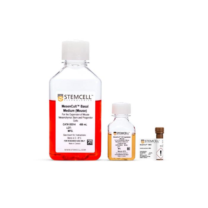
MesenCult™ 扩增试剂盒 (小鼠)

用于培养小鼠MSCs和MEFs
只有 %1
¥2,854.00

产品号 #(选择产品)

产品号 #05513_C

用于培养小鼠MSCs和MEFs

产品优势

  • 可实现P0代MSCs的快速扩增和高效富集
  • 优化用于小鼠骨髓、密质骨和脂肪来源的MSCs以及MEFs
  • 获得均质的小鼠MSC培养体系,同时保持三系分化潜力
  • 严格的原料筛选和质量控制,最大限度地减少了批次之间的差异性,并提高实验的可重复性。

产品组分包括

  • MesenCult™基础培养基(小鼠),450 mL
  • MesenCult™10X补充剂(小鼠),50 mL
  • MesenPure™,0.5 mL
You may notice that your reagent packaging looks slightly different from images displayed here or from previous orders. Due to pandemic-related plasticware shortages, we are temporarily using alternative bottles for this product. Rest assured that the products themselves and how you should use them have not changed.
专为您的实验方案打造的产品
要查看实验方案所需的所有配套产品,请参阅《实验方案与技术文档》

总览

MesenCult™ 扩增试剂盒 (小鼠)是标准的用于小鼠间充质间质细胞(MSCs,也称为间充质干细胞)和小鼠胚胎成纤维细胞(MEFs)培养的培养基。该试剂盒包括MesenCult™基础培养基(小鼠),MesenCult™10X补充剂(小鼠)和MesenPure™。MesenCult™扩增培养基已经过优化,可用于小鼠MSCs和MEFs的衍生和体外扩增,以及用于成纤维细胞集落形成单位(CFUF)的检测。该试剂盒使用小鼠品系C57BL/6的细胞进行优化。

为了促进MSCs和MEFs在细胞培养过程中的富集,只需在使用前添加MesenPure™以配置MesenCult™扩增完全培养基,而无需通过连续传代和频繁的培养基换液。虽然不是必需的,但强烈建议添加MesenPure™,因为与单独使用Mesenult™扩增完全培养基相比,所得到的MSC和MEF培养物更均质,并且表现出更强大的增殖、分化和集落形成能力。

注意:MesenCult™ 扩增培养基必须补充L-谷氨酰胺(产品号 #07100)。

分类
专用培养基
 
细胞类型
间充质干/祖细胞,鼠胚胎成纤维细胞
 
种属
小鼠
 
应用
细胞培养,克隆筛选,扩增
 
品牌
MesenCult
 
研究领域
药物发现和毒性检测
 

实验数据

Procedure Summary for Hematopoietic CFU Assays

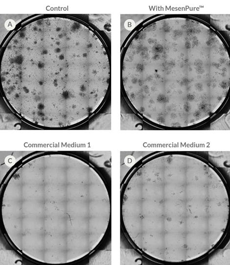
Figure 1. CFU-F Assay Comparing Mouse Bone Marrow (BM) MSCs Derived and Cultured in MesenCult™ Expansion Medium With and Without MesenPure™, and Other Commercially Available Media

Numerous CFU-F colonies were observed in cultures maintained in (A) MesenCult™ Expansion Medium (Control) and in (B) same medium containing MesenPure™. Few to no colony formation were observed when cultures were maintained in (C) Commercial Medium 1 or (D) Commercial Medium 2. Seeding density: 5x10^4 cells/cm^2.

Procedure Summary for Hematopoietic CFU Assays

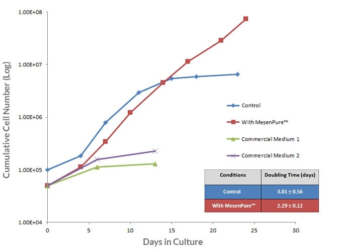
Figure 2. Long-Term Expansion of Mouse BM-Derived MSCs is Observed When Cells are Cultured in MesenCult™ Expansion Medium

Mouse BM MSCs, derived and cultured in MesenCult™ Expansion Medium (Control), show superior long-term expansion rate compared to Commercial Medium 1 and 2. The addition of MesenPure™ enriches for MSCs as early as passage 0 and further improves the expansion rate beyond passage 8. The doubling time of mouse MSCs cultured with or without MesenPure™ are 2.29 and 3.01, respectively. BM MSCs culture-expanded using the MesenCult™ Expansion Kit, with or without MesenPure™, were done under hypoxic conditions. BM MSCs culture-expanded in Commercial Medium 1 and 2 were culture-expanded under normoxic conditions as recommended by their protocols. Data shown from one representative experiment (n=3).

Procedure Summary for Hematopoietic CFU Assays

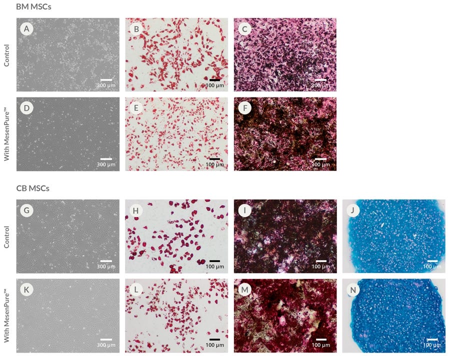
Figure 3. Mouse BM- and Compact Bone (CB)-Derived MSCs Culture-expanded in MesenCult™ Expansion Medium With or Without MesenPure™ Maintain Multi-Lineage Differentiation Potential

Enriched populations of MSCs were observed at earlier passages upon addition of MesenPure™, which showed increased and more dense differentiation than control cultures. (A) Mouse BM MSCs culture-expanded in MesenCult™ Expansion Medium (Control) differentiated into (B) adipocytes; and (C) osteoblasts. (D) Mouse BM-derived MSCs culture-expanded with MesenPure™ differentiated into (E) adipocytes; and (F) osteoblasts . Differentiation of mouse BM MSCs into chondrocytes is in progress. (G) Mouse CB MSCs culture-expanded in MesenCult™ Expansion Medium (Control) differentiated into (H) adipocytes, (I) osteoblasts and (J) chondrocytes. Adipose-derived mesenchymal stem and progenitor cells, and mouse embryonic fibroblasts (MEFs) were derived and culture-expanded using the MesenCult™ Expansion Kit. These cells were also differentiated towards the adipogenic and osteogenic lineages (data not shown). Adipocytes were stained with Oil Red O staining. Osteoblasts were stained with Alkaline phosphatase and silver nitrate (von Kossa). Chondrocytes were stained with Alcian Blue and Nuclear Fast Red. Images were taken at passage 2.

Procedure Summary for Hematopoietic CFU Assays

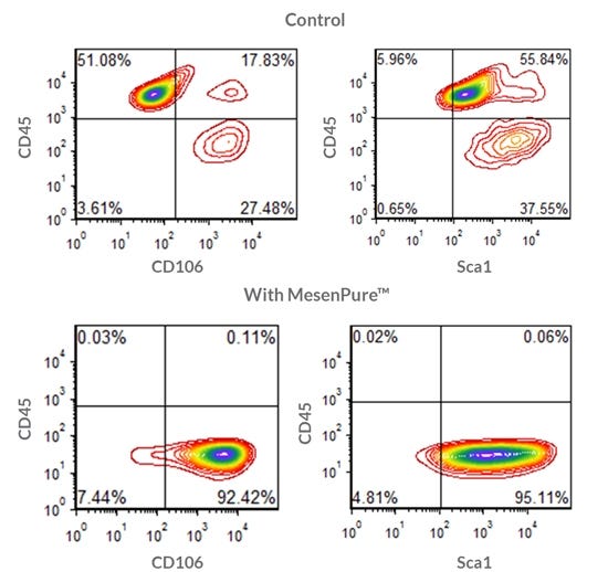
Figure 4. Flow Cytometric Analysis of Culture-Expanded Mouse BM-Derived MSCs Using the MesenCult™ Expansion Kit

Mouse BM MSCs were culture-expanded in MesenCult™ Expansion Medium (Control) or with MesenPure™. MSCs from passage 2 were stained for the mesenchymal surface markers, CD106 and Sca1, and the hematopoietic marker, CD45. Stained cells were then analyzed by flow cytometry. MSCs culture-expanded in Control medium show distinct populations of CD45+ hematopoietic cells and CD45- (CD106+ and Sca1+) MSCs. Upon addition of MesenPure™ to the Control Medium, an enriched and homogenous population of CD45- (CD106+ and Sca1+) MSCs are obtained.

产品说明书及文档

请在《产品说明书》中查找相关支持信息和使用说明,或浏览下方更多实验方案。

Document Type
Product Name
Catalog #
Lot #
Language
Catalog #
05513
Lot #
All
Language
English
Document Type
Safety Data Sheet 1
Catalog #
05513
Lot #
All
Language
English
Document Type
Safety Data Sheet 2
Catalog #
05513
Lot #
All
Language
English
Document Type
Safety Data Sheet 3
Catalog #
05513
Lot #
All
Language
English

应用领域

本产品专为以下研究领域设计,适用于工作流程中的高亮阶段。探索这些工作流程,了解更多我们为各研究领域提供的其他配套产品。

相关材料与文献

技术资料 (5)

文献 (16)

FXR Regulates Intestinal Cancer Stem Cell Proliferation. T. Fu et al. Cell 2019

Abstract

Increased levels of intestinal bile acids (BAs) are a risk factor for colorectal cancer (CRC). Here,we show that the convergence of dietary factors (high-fat diet) and dysregulated WNT signaling (APC mutation) alters BA profiles to drive malignant transformations in Lgr5-expressing (Lgr5+) cancer stem cells and promote an adenoma-to-adenocarcinoma progression. Mechanistically,we show that BAs that antagonize intestinal farnesoid X receptor (FXR) function,including tauro-$\beta$-muricholic acid (T-$\beta$MCA) and deoxycholic acid (DCA),induce proliferation and DNA damage in Lgr5+ cells. Conversely,selective activation of intestinal FXR can restrict abnormal Lgr5+ cell growth and curtail CRC progression. This unexpected role for FXR in coordinating intestinal self-renewal with BA levels implicates FXR as a potential therapeutic target for CRC.
Mesenchymal Stromal Cells Modulate Corneal Alloimmunity via Secretion of Hepatocyte Growth Factor. S. K. Mittal et al. Stem cells translational medicine 2019 jun

Abstract

Mesenchymal stromal cells (MSCs) are multipotent stem cells that participate in tissue repair and posses considerable immunomodulatory potential. MSCs have been shown to promote allograft survival,yet the mechanisms behind this phenomenon have not been fully defined. Here,we investigate the capacity of MSCs to suppress the allogeneic immune response by secreting the pleiotropic molecule hepatocyte growth factor (HGF). Using an in vivo mouse model of corneal transplantation,we report that MSCs promote graft survival in an HGF-dependent manner. Moreover,our data indicate that topically administered recombinant HGF (1) suppresses antigen-presenting cell maturation in draining lymphoid tissue,(2) limits T-helper type-1 cell generation,(3) decreases inflammatory cell infiltration into grafted tissue,and (4) is itself sufficient to promote transplant survival. These findings have potential translational implications for the development of HGF-based therapeutics. Stem Cells Translational Medicine 2019.
Carnosine Supplementation Mitigates the Deleterious Effects of Particulate Matter Exposure in Mice. W. Abplanalp et al. Journal of the American Heart Association 2019 jul

Abstract

Background Exposure to fine airborne particulate matter ( PM 2.5) induces quantitative and qualitative defects in bone marrow-derived endothelial progenitor cells of mice,and similar outcomes in humans may contribute to vascular dysfunction and the cardiovascular morbidity and mortality associated with PM 2.5 exposure. Nevertheless,mechanisms underlying the pervasive effects of PM 2.5 are unclear and effective interventional strategies to mitigate against PM 2.5 toxicity are lacking. Furthermore,whether PM 2.5 exposure affects other types of bone marrow stem cells leading to additional hematological or immunological dysfunction is not clear. Methods and Results Mice given normal drinking water or that supplemented with carnosine,a naturally occurring,nucleophilic di-peptide that binds reactive aldehydes,were exposed to filtered air or concentrated ambient particles. Mice drinking normal water and exposed to concentrated ambient particles demonstrated a depletion of bone marrow hematopoietic stem cells but no change in mesenchymal stem cells. However,HSC depletion was significantly attenuated when the mice were placed on drinking water containing carnosine. Carnosine supplementation also increased the levels of carnosine-propanal conjugates in the urine of CAPs-exposed mice and prevented the concentrated ambient particles-induced dysfunction of endothelial progenitor cells as assessed by in vitro and in vivo assays. Conclusions These results suggest that exposure to PM 2.5 has pervasive effects on different bone marrow stem cell populations and that PM 2.5-induced hematopoietic stem cells depletion,endothelial progenitor cell dysfunction,and defects in vascular repair can be mitigated by excess carnosine. Carnosine supplementation may be a viable approach for preventing PM 2.5-induced immune dysfunction and cardiovascular injury in humans.

更多信息

更多信息
物种 小鼠
质量保证:

产品仅供研究使用,不用于针对人或动物的诊断或治疗。 欲获悉更多关于STEMCELL的质控信息,请访问 STEMCELL.CN/COMPLIANCE.
Copyright © 2026 by STEMCELL Technologies. All rights reserved.

在线联系