若您需要咨询产品或有任何技术问题,请通过官方电话 400 885 9050 或邮箱 info.cn@stemcell.com 与我们联系。

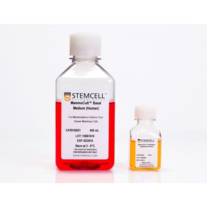
MammoCult™ 人源培养基套装

用于人乳腺球和肿瘤球的培养
只有 %1
¥4,934.00

产品号 #(选择产品)

产品号 #05620

 用于人乳腺球和肿瘤球的培养

产品组分包括

  • MammoCult™基础培养基(人源),450 mL
  • MammoCult™扩增补充剂(人源),50 mL
专为您的实验方案打造的产品
要查看实验方案所需的所有配套产品,请参阅《实验方案与技术文档》

总览

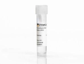
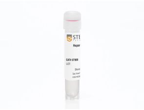
MammoCult™培养基(人源)是一种无血清、不含雌激素和孕酮的培养基,专为培养正常人原代乳腺组织来源的乳腺球和人乳腺癌细胞系来源的肿瘤球而优化。配制完整的MammoCult™培养基还需使用氢化可的松储备液(产品号#07925)和肝素溶液(产品号#07980)。

分类
专用培养基
 
细胞类型
癌细胞及细胞系,乳腺细胞
 
种属

 
应用
细胞培养,培养,球状体培养
 
品牌
MammoCult
 
研究领域
癌症,上皮细胞研究
 
制剂类别
无血清
 

实验数据

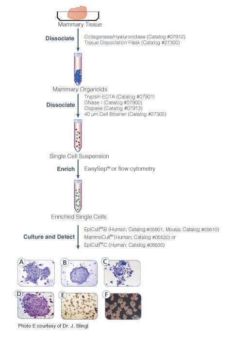
Protocol for isolation and identification of human and mouse mammary epithelial progenitor cells

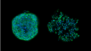
Figure 1. Protocol for Isolation and Identification of Human and Mouse Mammary Epithelial Progenitor Cells

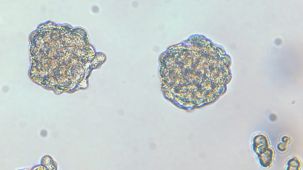
Phase contrast photographs of (A) a pure human myoepithelial cell colony, (B) a pure human luminal cell colony, and (C) a mixed human colony. (D) is a mouse colony. Unlike human mammary CFC colonies, subtypes of mouse mammary epithelial cell colonies are not easily identifiable. All colonies were cultured in either EpiCult™-B (Human: Catalog #05601) or EpiCult™-B (Mouse:Catalog #5610) in the presence of an irradiated NIH 3T3 feeder layer. Colonies were visualized by staining with Wright"s Giemsa. (E) is a picture of mammospheres obtained from primary human mammary epithelial cells and (F) is an image of tumorspheres obtained from MCF7 human breast cancer cell line.

产品说明书及文档

请在《产品说明书》中查找相关支持信息和使用说明,或浏览下方更多实验方案。

Document Type
Product Name
Catalog #
Lot #
Language
Catalog #
05620
Lot #
All
Language
English
Document Type
Safety Data Sheet 1
Catalog #
05620
Lot #
All
Language
English
Document Type
Safety Data Sheet 2
Catalog #
05620
Lot #
All
Language
English

应用领域

本产品专为以下研究领域设计,适用于工作流程中的高亮阶段。探索这些工作流程,了解更多我们为各研究领域提供的其他配套产品。

相关材料与文献

技术资料 (12)

文献 (90)

Generation of a functional mammary gland from a single stem cell. Shackleton M et al. Nature 2006 JAN

Abstract

The existence of mammary stem cells (MaSCs) has been postulated from evidence that the mammary gland can be regenerated by transplantation of epithelial fragments in mice. Interest in MaSCs has been further stimulated by their potential role in breast tumorigenesis. However,the identity and purification of MaSCs has proved elusive owing to the lack of defined markers. We isolated discrete populations of mouse mammary cells on the basis of cell-surface markers and identified a subpopulation (Lin-CD29hiCD24+) that is highly enriched for MaSCs by transplantation. Here we show that a single cell,marked with a LacZ transgene,can reconstitute a complete mammary gland in vivo. The transplanted cell contributed to both the luminal and myoepithelial lineages and generated functional lobuloalveolar units during pregnancy. The self-renewing capacity of these cells was demonstrated by serial transplantation of clonal outgrowths. In support of a potential role for MaSCs in breast cancer,the stem-cell-enriched subpopulation was expanded in premalignant mammary tissue from MMTV-wnt-1 mice and contained a higher number of MaSCs. Our data establish that single cells within the Lin-CD29hiCD24+ population are multipotent and self-renewing,properties that define them as MaSCs.
Novel cell culture technique for primary ductal carcinoma in situ: role of Notch and epidermal growth factor receptor signaling pathways. Farnie G et al. Journal of the National Cancer Institute 2007 APR

Abstract

BACKGROUND The epidermal growth factor receptor (EGFR) and Notch signaling pathways have been implicated in self-renewal of normal breast stem cells. We investigated the involvement of these signaling pathways in ductal carcinoma in situ (DCIS) of the breast. METHODS Samples of normal breast tissue (n = 15),pure DCIS tissue of varying grades (n = 35),and DCIS tissue surrounding an invasive cancer (n = 7) were used for nonadherent (i.e.,mammosphere) culture. Mammosphere cultures were treated at day 0 with gefitinib (an EGFR inhibitor),DAPT (N-[N-(3,5-difluorophenacetyl-L-alanyl)]-S-phenylglycine t-butyl ester) (a gamma-secretase inhibitor),or Notch 4-neutralizing antibody. Mammosphere-forming efficiency (MFE) was calculated by dividing the number of mammospheres of 60 microm or more formed by the number of single cells seeded and is expressed as a percentage. The Notch 1 intracellular domain (NICD) was detected immunohistochemically in paraffin-embedded DCIS tissue from 50 patients with at least 60 months of follow-up. All statistical tests were two-sided. RESULTS DCIS had a greater MFE than normal breast tissue (1.5% versus 0.5%,difference = 1%,95% confidence interval [CI] = 0.62% to 1.25%,Ptextless.001). High-grade DCIS had a greater MFE than low-grade DCIS (1.6% versus 1.09%,difference = 0.51%,95% CI = 0.07% to 0.94%,P = .01). The MFE of high-grade DCIS treated with gefitinib in the absence of exogenous EGF was lower than that of high-grade DCIS treated with mammosphere medium lacking gefitinib and exogenous EGF (0.56% versus 1.36%,difference 0.8%,95% CI = 0.33% to 1.4%,P = .004). Increased Notch signaling as detected by NICD staining was associated with recurrence at 5 years (P = .012). DCIS MFE was reduced when Notch signaling was inhibited using either DAPT (0.89% versus 0.51%,difference = 0.38%,95% CI = 0.2% to 0.6%,Ptextless.001) or a Notch 4-neutralizing antibody (0.97% versus 0.2%,difference = 0.77%,95% CI = 0.52% to 1.0%,Ptextless.001). CONCLUSION We describe a novel primary culture technique for DCIS. Inhibition of the EGFR or Notch signaling pathways reduced DCIS MFE.
Distinct expression levels and patterns of stem cell marker, aldehyde dehydrogenase isoform 1 (ALDH1), in human epithelial cancers. Deng S et al. PloS one 2010 JAN

Abstract

Aldehyde dehydrogenase isoform 1 (ALDH1) has been proved useful for the identification of cancer stem cells. However,our knowledge of the expression and activity of ALDH1 in common epithelial cancers and their corresponding normal tissues is still largely absent. Therefore,we characterized ALDH1 expression in 24 types of normal tissues and a large collection of epithelial tumor specimens (six cancer types,n = 792) by immunohistochemical staining. Using the ALDEFUOR assay,ALDH1 activity was also examined in 16 primary tumor specimens and 43 established epithelial cancer cell lines. In addition,an ovarian cancer transgenic mouse model and 7 murine ovarian cancer cell lines were analyzed. We found that the expression levels and patterns of ALDH1 in epithelial cancers are remarkably distinct,and they correlate with their corresponding normal tissues. ALDH1 protein expression levels are positively correlated with ALDH1 enzymatic activity measured by ALDEFLUOR assay. Long-term in vitro culture doesn't significantly affect ALDH1 activity in epithelial tumor cells. Consistent with research on other cancers,we found that high ALDH1 expression is significantly associated with poor clinical outcomes in serous ovarian cancer patients (n = 439,p = 0.0036). Finally,ALDH(br) tumor cells exhibit cancer stem cell properties and are resistant to chemotherapy. As a novel cancer stem cell marker,ALDH1 can be used for tumors whose corresponding normal tissues express ALDH1 in relatively restricted or limited levels such as breast,lung,ovarian or colon cancer.

更多信息

更多信息
物种
配方 无血清
质量保证:

产品仅供研究使用,不用于针对人或动物的诊断或治疗。 欲获悉更多关于STEMCELL的质控信息,请访问 STEMCELL.CN/COMPLIANCE.
Copyright © 2026 by STEMCELL Technologies. All rights reserved.

在线联系