若您需要咨询产品或有任何技术问题,请通过官方电话 400 885 9050 或邮箱 info.cn@stemcell.com 与我们联系。

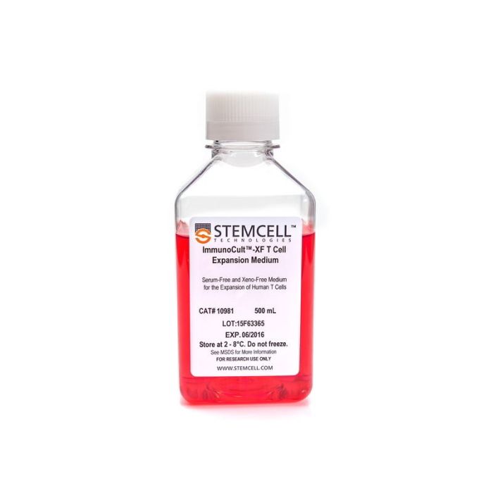
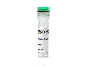
ImmunoCult™ XF 人T细胞扩增培养基

用于T细胞扩增的无血清和无异种成分培养基
只有 %1
¥1,750.00

产品号 #(选择产品)

产品号 #10981_C

用于T细胞扩增的无血清和无异种成分培养基

产品优势

  • 无需在培养基中添加血清
  • 支持稳定的T细胞扩增,培养10 - 12天后仍具有较高的活率
  • 扩增的T细胞再刺激时能够产生包括IFN-γ和IL-4在内的细胞因子
  • 与ImmunoCult™人T细胞激活剂(产品号#10970或#10971)搭配使用,用于T细胞的无磁珠活化

总览

Immunocult™-XF T细胞扩增培养基是一种无血清和无异种成分培养基,用于体外培养和扩增外周血中分选的T细胞。适合T细胞的生长和扩增的重组细胞因子尚未添加到ImmunoCult™-XF T细胞扩增培养基中,以便用户灵活地制备满足其要求的完全培养基。
本产品仅用于为科研,如果您需要适合细胞治疗生产级别的试剂,推荐ImmunoCult™-XF(产品号#100-0956),该产品生产满足cGMP级相关规定,可用于临床应用。

分类
专用培养基
 
细胞类型
T 细胞,T 细胞,CD4+,T 细胞,CD8+
 
种属
人,小鼠
 
应用
细胞培养,扩增
 
品牌
ImmunoCult
 
研究领域
细胞疗法开发,药物发现和毒性检测,免疫学
 
制剂类别
无血清,无异源
 

实验数据

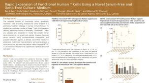
ImmunoCult™-XF T Cell Expansion Medium Supports Faster T Cell Expansion Than Other Serum-Free and Serum-Supplemented Media

Figure 1. ImmunoCult™-XF T Cell Expansion Medium Supports Faster T Cell Expansion Than Other Serum-Free and Serum-Supplemented Media

T cells were isolated from human peripheral blood samples using the EasySep™ Human T Cell Isolation Kit (Catalog #17951), stimulated with ImmunoCult™ Human CD3/CD28/CD2 T Cell Activator (Catalog #10970), and cultured in ImmunoCult™-XF T Cell Expansion Medium supplemented with rhIL-2. T cells were stimulated with ImmunoCult™ Human CD3/CD28/CD2 T Cell Activator on day 0 and every 7 to 8 days for the duration of the culture. T cells were analyzed on days 4, 7, 8, 10, 11, 14, 18, and 21 for fold expansion relative to the initial cell seeding density. Compared to all competitor media tested, ImmunoCult™-XF T Cell Expansion Medium showed significantly higher expansion of total T cells. Competitors 1 to 4 include, in no particular order, X-VIVO™ 15 (Lonza), AIM V® Medium (Life Tech), CellGro® DC Medium (CellGenix), and RPMI 1640 + serum. Each data point represents the mean fold expansion ± S.E.M. at the specified time points (p<0.05 for ImmunoCult™-XF versus all media for days 8, 11, 14, 18, and 21, tested using two-tailed, paired t-test with unequal variance, n = 6 to 19 donors). The average fold expansion of T cells in ImmunoCult™-XF T Cell Expansion Medium were 15-fold on day 7, 80-fold on day 10, 450-fold on day 14, and 4,000-fold on day 21.

ImmunoCult™-XF T Cell Expansion Medium Supports Greater T Cell Expansion Than Other Serum-Free and Serum-Supplemented Media

Figure 2. ImmunoCult™-XF T Cell Expansion Medium Supports Greater T Cell Expansion Than Other Serum-Free and Serum-Supplemented Media

T cells were isolated from human peripheral blood samples using the EasySep™ Human T Cell Isolation Kit (Catalog #17951), stimulated with ImmunoCult™ Human CD3/CD28/CD2 T Cell Activator (Catalog #10970), and cultured in (A) ImmunoCult™-XF T Cell Expansion Medium or serum-free competitor media with rhIL-2 in three replicate cultures per donor, or cultured in (B) ImmunoCult™-XF T Cell Expansion Medium or serum-supplemented competitor media with rhIL-2 in three replicate cultures per donor. T cells were stimulated with ImmunoCult™ Human CD3/CD28/CD2 T Cell Activator on day 0 and every 7 to 8 days for the duration of the culture. T cells were analyzed on day 21 for fold expansion relative to the initial cell seeding density.
(A) Compared to all serum-free competitor media tested, ImmunoCult™-XF T Cell Expansion Medium showed significantly higher expansion of total T cells. Competitors 1 to 6 represent serum-free competitor media, which include, in no particular order, X-VIVO™ 15 (Lonza), AIM V® Medium (Life Tech), CellGro® DC Medium (CellGenix), CTS™ OpTmizer™ T Cell Expansion SFM (Life Tech), TexMACS™ Medium (Miltenyi), and PRIME-XV® T Cell Expansion XSFM (Irvine Scientific). Each column with error bars represents the mean ± S.E.M. (p<5x10-13 for ImmunoCult™-XF T Cell Expansion Medium versus all other serum-free media, tested using the linear mixed effect model with linear regression, n = 4 to 19 donors).
(B) Compared to all serum-supplemented competitor media tested, ImmunoCult™-XF T Cell Expansion Medium showed similar or significantly higher expansion of total T cells. Competitors 1 to 4 represent serum-supplemented competitor media, which include, in no particular order, X-VIVO™ 15 + serum, CTS™ OpTmizer™ T Cell Expansion SFM + serum, RPMI 1640 + serum, and IMDM + serum. Each column with error bars represents the mean ± S.E.M. (p<0.0006 for ImmunoCult™-XF T Cell Expansion Medium versus all other serum-supplemented media except for Competitor 4, tested using the linear mixed effect model with linear regression, n = 1 to 19 donors).

T Cells Expanded in ImmunoCult™-XF T Cell Expansion Medium Show Similar Proportions of CD4+ and CD8+ Cells as T Cells at the Start of Culture

Figure 3. T Cells Expanded in ImmunoCult™-XF T Cell Expansion Medium Show Similar Proportions of CD4+ and CD8+ Cells as T Cells at the Start of Culture

T cells were isolated from human peripheral blood samples using the EasySep™ Human T Cell Isolation Kit (Catalog #17951), stimulated with ImmunoCult™ Human CD3/CD28/CD2 T Cell Activator (Catalog #10970), and cultured in ImmunoCult™-XF T Cell Expansion Medium supplemented with rhIL-2. T cells were stimulated with ImmunoCult™ Human CD3/CD28/CD2 T Cell Activator on day 0 and every 7 to 8 days for the duration of the culture. On day 0 and day 21, T cells were harvested and analyzed for (A) CD4+ and (B) CD8+ expression. Each column with error bars represents the mean ± S.E.M. (n = 24 donors for day 0 and n = 19 donors for day 21).

T Cells Expanded in ImmunoCult™-XF T Cell Expansion Medium Produce Intracellular IFN-gamma and IL-4

Figure 4. T Cells Expanded in ImmunoCult™-XF T Cell Expansion Medium Produce Intracellular IFN-gamma and IL-4

T cells were isolated from human peripheral blood samples using the EasySep™ Human T Cell Isolation Kit (Catalog #17951), stimulated with ImmunoCult™ Human CD3/CD28/CD2 T Cell Activator (Catalog #10970), and cultured in ImmunoCult™-XF T Cell Expansion Medium supplemented with rhIL-2. T cells were stimulated with ImmunoCult™ Human CD3/CD28/CD2 T Cell Activator on day 0 and every 7 to 8 days for the duration of the culture. On day 21, T cells were harvested and analyzed for intracellular IFN-gamma and IL-4 after stimulation with PMA and ionomycin for 4 hours and with Brefeldin A for 2 hours. The production of IFN-gamma and IL-4 in CD3+, CD3+CD4+CD8-, and CD3+CD4-CD8+ cells were determined. Each stacked column with error bars represents the mean ± S.E.M. (n = 9 donors).

产品说明书及文档

请在《产品说明书》中查找相关支持信息和使用说明,或浏览下方更多实验方案。

Document Type
Product Name
Catalog #
Lot #
Language
Catalog #
10981
Lot #
All
Language
English
Document Type
Safety Data Sheet
Catalog #
10981
Lot #
All
Language
English

应用领域

本产品专为以下研究领域设计,适用于工作流程中的高亮阶段。探索这些工作流程,了解更多我们为各研究领域提供的其他配套产品。

相关材料与文献

技术资料 (35)

文献 (37)

Tuning ITAM multiplicity on T cell receptors can control potency and selectivity to ligand density. J. R. James Science signaling 2018 MAY

Abstract

The T cell antigen receptor (TCR) recognizes peptides from pathogenic proteins bound in the major histocompatibility complex (MHC). To convert this binding event into downstream signaling,the TCR complex contains immunoreceptor tyrosine-based activation motifs (ITAMs) that act as docking sites for the cytoplasmic tyrosine kinase ZAP-70. Unique among antigen receptors,the TCR complex uses 10 ITAMs to transduce peptide-MHC binding to the cell interior. Using synthetic,drug-inducible receptor-ligand pairs,it was found that greater ITAM multiplicity primarily enhanced the efficiency with which ligand binding was converted into an intracellular signal. This manifested as an increase in the fraction of cells that became activated in response to antigen,and a more synchronous initiation of TCR-proximal signaling,rather than direct amplification of the intracellular signals. Exploiting these findings,the potency and selectivity of chimeric antigen receptors targeted against cancer were substantially enhanced by modulating the number of encoded ITAMs.
NoxO1 Controls Proliferation of Colon Epithelial Cells. F. Moll et al. Frontiers in immunology 2018

Abstract

Aim Reactive oxygen species (ROS) produced by enzymes of the NADPH oxidase family serve as second messengers for cellular signaling. Processes such as differentiation and proliferation are regulated by NADPH oxidases. In the intestine,due to the exceedingly fast and constant renewal of the epithelium both processes have to be highly controlled and balanced. Nox1 is the major NADPH oxidase expressed in the gut,and its function is regulated by cytosolic subunits such as NoxO1. We hypothesize that the NoxO1-controlled activity of Nox1 contributes to a proper epithelial homeostasis and renewal in the gut. Results NoxO1 is highly expressed in the colon. Knockout of NoxO1 reduces the production of superoxide in colon crypts and is not subsidized by an elevated expression of its homolog p47phox. Knockout of NoxO1 increases the proliferative capacity and prevents apoptosis of colon epithelial cells. In mouse models of dextran sulfate sodium (DSS)-induced colitis and azoxymethane/DSS induced colon cancer,NoxO1 has a protective role and may influence the population of natural killer cells. Conclusion NoxO1 affects colon epithelium homeostasis and prevents inflammation.
Translational control of tumor immune escape via the eIF4F-STAT1-PD-L1 axis in melanoma. M. Cerezo et al. Nature medicine 2018 OCT

Abstract

Preventing the immune escape of tumor cells by blocking inhibitory checkpoints,such as the interaction between programmed death ligand-1 (PD-L1) and programmed death-1 (PD-1) receptor,is a powerful anticancer approach. However,many patients do not respond to checkpoint blockade. Tumor PD-L1 expression is a potential efficacy biomarker,but the complex mechanisms underlying its regulation are not completely understood. Here,we show that the eukaryotic translation initiation complex,eIF4F,which binds the 5' cap of mRNAs,regulates the surface expression of interferon-$\gamma$-induced PD-L1 on cancer cells by regulating translation of the mRNA encoding the signal transducer and activator of transcription 1 (STAT1) transcription factor. eIF4F complex formation correlates with response to immunotherapy in human melanoma. Pharmacological inhibition of eIF4A,the RNA helicase component of eIF4F,elicits powerful antitumor immune-mediated effects via PD-L1 downregulation. Thus,eIF4A inhibitors,in development as anticancer drugs,may also act as cancer immunotherapies.

更多信息

更多信息
物种 人, 小鼠
配方 无血清
质量保证:

产品仅供研究使用,不用于针对人或动物的诊断或治疗。 欲获悉更多关于STEMCELL的质控信息,请访问 STEMCELL.CN/COMPLIANCE.
Copyright © 2026 by STEMCELL Technologies. All rights reserved.

在线联系