若您需要咨询产品或有任何技术问题,请通过官方电话 400 885 9050 或邮箱 info.cn@stemcell.com 与我们联系。

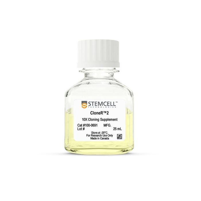
CloneR™2

用于提高人胚胎干细胞和诱导多能干细胞在单细胞工作流程中存活率的添加物

只有 %1
¥2,440.00

产品号 #(选择产品)

产品号 #100-0691_C

用于提高人胚胎干细胞和诱导多能干细胞在单细胞工作流程中存活率的添加物

产品优势

  • 更多克隆,提前准备。克隆效率提高,使克隆体更早准备好进行筛选。
  • 稳定高效且一致的克隆表现。在不同的培养系统和细胞系中均表现出高性能。
  • 增强的存活率。在各种密度下以及经历电转或冻存后复苏等高应激事件后,接种效率提高。
  • 直接进行单细胞操作。无需单细胞传代适应阶段。

总览

使用 CloneR™2可生成可保持基因组完整性和下游分化潜能的人多能干细胞 (hPSC) 克隆系。可在各种高应激条件下(包括高密度或低密度接种)显著提高人胚胎干细胞(ES)和诱导多能干细胞(iPS)的克隆效率和存活率。

在您的基因编辑流程中,添加 CloneR™2 可提升 ES 和 iPS 细胞电转后以及单细胞沉降(Single-cell deposition)的存活率(参见下文数据)。该添加物还可促进低密度接种(<25 个细胞/cm²)或 FACS 单细胞分选(1 个细胞/孔)获得的克隆细胞的存活和扩增,无需进行单细胞适应过程。同时,CloneR™2 也适用于在较高密度铺板的单细胞存活,包括诸如冻存复苏、或分化前建立单层细胞等应激条件下的应用。

CloneR™2 是一种成分明确、无血清的添加物,可与 TeSR™ ES 和 iPS 细胞培养基兼容使用,并适用于您选择的细胞培养基质。

分类
添加剂
 
细胞类型
多能干细胞
 
种属

 
应用
细胞培养
 
品牌
CloneR
 
研究领域
细胞系制备,疾病建模,干细胞生物学
 
制剂类别
无血清
 

实验数据

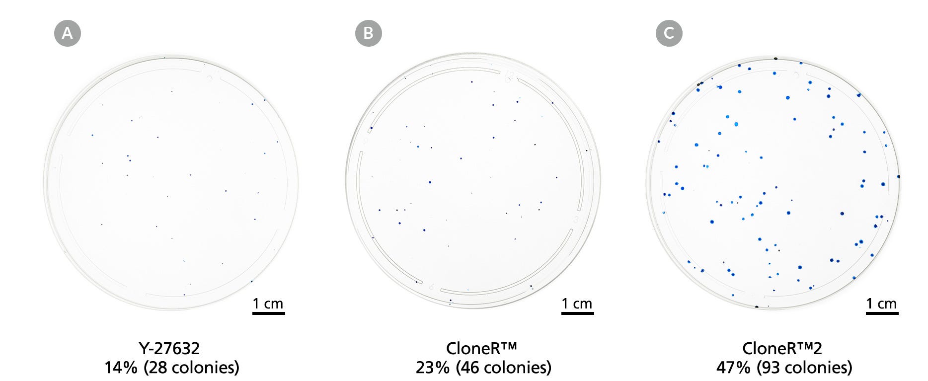
Three 10 cm dishes showing increased numbers of colonies from Y-27632 compound to CloneR™ to CloneR™2

Figure 1. CloneR™ and CloneR™2 Supplements Improve Cloning Efficiency and Colony Size

hPSCs display a considerable increase in cloning efficiency when cloned using (B) CloneR™ compared to using (A) Y-27632 compound. (C) CloneR™2 further improves cloning efficiency and increases colony size when compared to either Y-27632 compound or CloneR™. Shown are examples of H9 hESCs in 10-cm dishes, plated at 200 cells per dish (~4 cells/cm²) in mTeSR™ Plus on Vitronectin XF™.

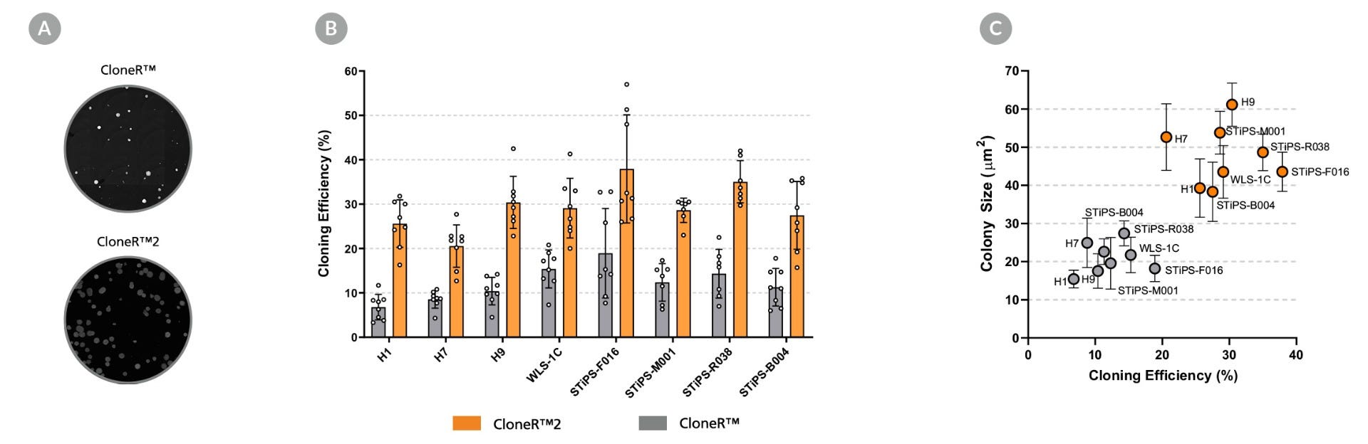
Images of colonies next to two graphs showing increased cloning efficiency and colony size in CloneR™2 across cell lines.

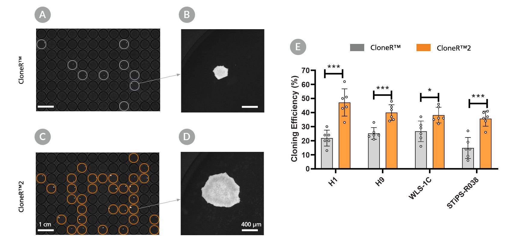
Figure 2. Use of CloneR™2 Enables Improved Cloning Efficiency and Larger Colonies When Compared to Use of CloneR™

(A) Representative images of 200 cells (H9 cell line) in 12-well plates grown in mTeSR™1 on Vitronectin XF™ at day 8 after seeding. Three hES (H1, H7 and H9) and 5 hiPS (WLS-1C, STiPS-F016, STiPS-M001, STiPS-R038 and STiPS-B004) cell lines were seeded at clonal density (50 cells/cm²) on Vitronectin XF™, in mTeSR™1 supplemented with CloneR™ or CloneR™2. mTeSR™1 supplemented with CloneR™2 increases (B) cloning efficiency and (C) colony size of hPSCs when compared with mTeSR™1 supplemented with CloneR™. Each data point in (B) represents an average of 3 technical replicates, with at least 7 biological replicates (n) per cell line.

Images of 96 well plates of cells with more colonies in CloneR™2 and summarized cloning efficiencies shown in a bar graph.

Figure 3. CloneR™2 Improves Clonal Generation Following Single Cell Deposition

Single-cell deposition is the gold standard of cloning; it relies on a FACS-based method that typically results in low cloning efficiency. Clones generated in mTeSR™ Plus supplemented with CloneR™2 demonstrate significantly improved (E) cloning efficiency and (B, D) colony size when compared to clones generated in mTeSR™ Plus supplemented with CloneR™. (A, C) Representative images of H9 hESCs cultured for eight days plated on Vitronectin XF™. Across four cell lines tested, CloneR™ and CloneR™2 had an average cloning efficiency of 21.2 ± 5.6% and 38.6 ± 6.0%, respectively, with at least 3 biological replicates per cell line. * denotes p < 0.05; *** denotes p < 0.001 by unpaired t-tests.

Graph of seeding efficiency of multiple cell lines in Y-27632 compared to CloneR™2.

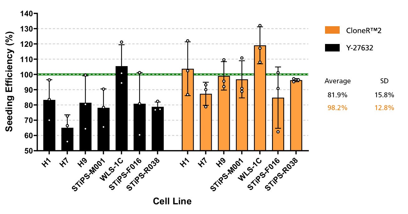
Figure 4. CloneR™2 Improves Seeding Efficiency at High Density

CloneR™2 improves single-cell seeding efficiency when used as a supplement in media for the first 24 hours of culture, compared to using Y-27632 as a supplement. 5.0x10⁵ cells were seeded in 12-well plates coated with Corning® Matrigel® in mTeSR™ Plus supplemented with CloneR™2 or Y-27632. Cultures were analyzed 24 hours post-seeding. The use of CloneR™2 resulted in an average seeding efficiency of 98.2 ± 12.8% compared to the use of Y-27632, which resulted in an average seeding efficiency of 81.9 ± 15.8%, across all cell lines (n = 3 replicates per line).

Graph of increased expansion of hPSCs when plated in CloneR™2 compared to Y-27632

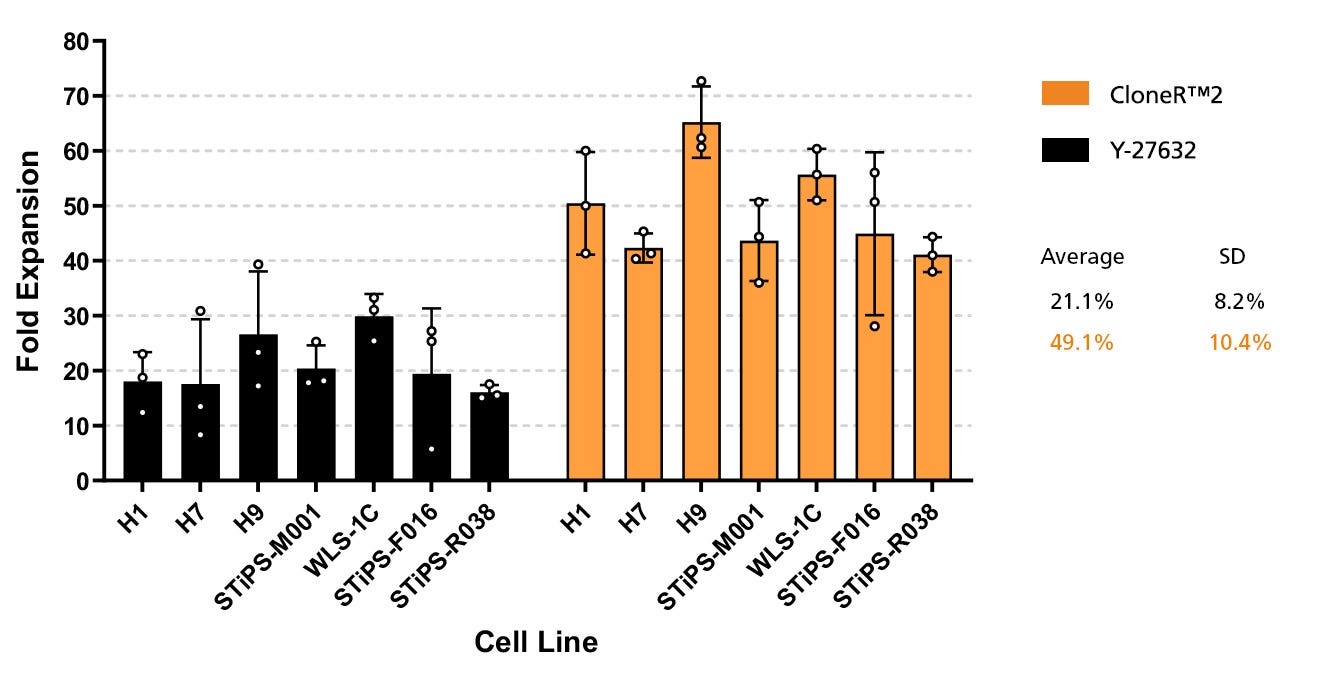
Figure 5. hPSCs Plated in CloneR™2 Show Increased Expansion

When used as a seeding supplement during single-cell passaging, CloneR™2 improves cell expansion when compared to using Y-27632. 3.0x10⁴ cells were seeded in 12-well plates coated with Corning® Matrigel® in mTeSR™ Plus supplemented with CloneR™2 or Y-27632. After 24 hours, the cultures were maintained in complete media (without a cloning supplement) and analyzed on day 5. CloneR™2 resulted in an average expansion of 49.1 ± 10.4 compared to Y-27632, which resulted in a lower average expansion of 21.1 ± 8.2, across all cell lines (n = 3 replicates per line).

Four graphs representing different cell lines for improved hPSC expansion in CloneR™2 following electroporation.

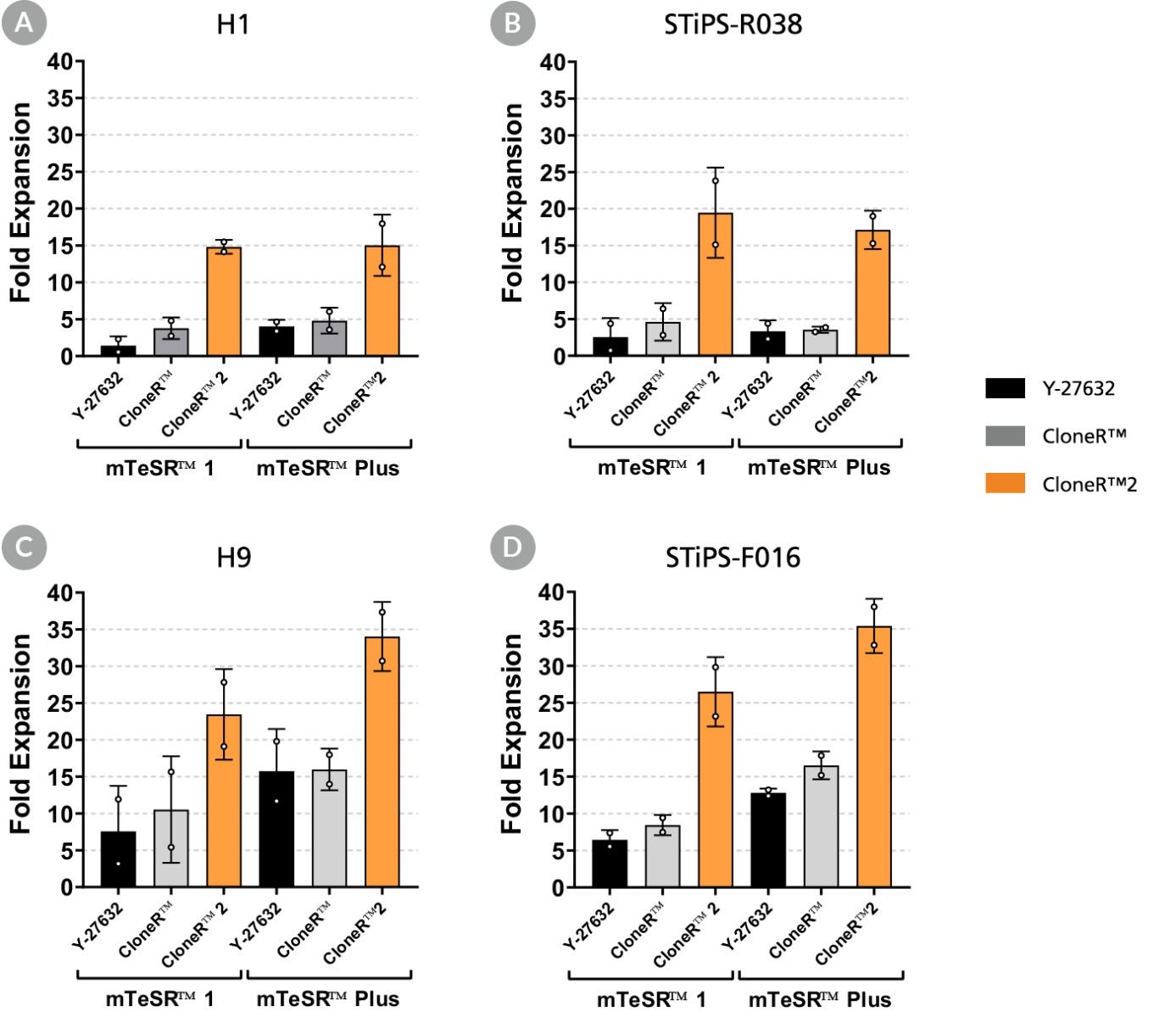
Figure 6. CloneR™2 Improves Recovery of hPSCs Following Electroporation

CloneR™2 can also be used as a survival supplement in gene-editing workflows that require electroporation. Four cell lines were electroporated, then plated in mTeSR™1 and mTeSR™ Plus containing Y-27632, CloneR™, or CloneR™2. Cultures were maintained in complete TeSR™ media (without cloning supplement) after 24 hours and analyzed after 5 days. In all 4 cell lines, (panels A-D) CloneR™2 dramatically improved cell survival and expansion when used as a supplement in the first 24 hours immediately following electroporation compared to both Y-27632 and CloneR™ (n = 2 replicates per cell line).

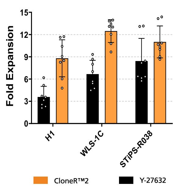
Bar graph showing improved post-thaw recovery of hPSCs in multiple cell lines when using CloneR™2

Figure 7. CloneR™2 Improves Post-Thaw Recovery of hPSCs

Thawing cryopreserved cells can result in low expansion or loss of the culture within the first passage. Using CloneR™2 as a seeding supplement within the first 24 hours of thawing cells ameliorates this effect, improving post-thaw recovery of hPSCs. Three cell lines were frozen as single cells, then thawed into mTeSR™ Plus containing Y-27632 or CloneR™2 on Matrigel®. Cultures were maintained in complete mTeSR™ Plus (without cloning supplement) after 24 hours, and analyzed on day 4 or day 5. CloneR™2 improves the fold-expansion across all cell lines tested when compared to Y-27632, with at least 7 replicates (n) per cell line.

产品说明书及文档

请在《产品说明书》中查找相关支持信息和使用说明,或浏览下方更多实验方案。

Document Type
Product Name
Catalog #
Lot #
Language
Product Name
CloneR™2
Catalog #
100-0691
Lot #
All
Language
English
Document Type
Safety Data Sheet
Product Name
CloneR™2
Catalog #
100-0691
Lot #
All
Language
English

应用领域

本产品专为以下研究领域设计,适用于工作流程中的高亮阶段。探索这些工作流程,了解更多我们为各研究领域提供的其他配套产品。

相关材料与文献

技术资料 (16)

文献 (10)

Dynein and dynactin move long-range but are delivered separately to the axon tip The Journal of Cell Biology 2024 Feb

Abstract

Fellows et al. report that individual dynein motors can move the entire axon length during retrograde transport. They find factors LIS1 and NDEL1,needed for transport initiation,also move with cargos. In the anterograde direction,dynein and its cofactor dynactin are transported separately,keeping them apart until required. Axonal transport is essential for neuronal survival. This is driven by microtubule motors including dynein,which transports cargo from the axon tip back to the cell body. This function requires its cofactor dynactin and regulators LIS1 and NDEL1. Due to difficulties imaging dynein at a single-molecule level,it is unclear how this motor and its regulators coordinate transport along the length of the axon. Here,we use a neuron-inducible human stem cell line (NGN2-OPTi-OX) to endogenously tag dynein components and visualize them at a near-single molecule regime. In the retrograde direction,we find that dynein and dynactin can move the entire length of the axon (>500 µm). Furthermore,LIS1 and NDEL1 also undergo long-distance movement,despite being mainly implicated with the initiation of dynein transport. Intriguingly,in the anterograde direction,dynein/LIS1 moves faster than dynactin/NDEL1,consistent with transport on different cargos. Therefore,neurons ensure efficient transport by holding dynein/dynactin on cargos over long distances but keeping them separate until required.
The immunopathological landscape of human pre-TCRα deficiency: from rare to common variants M. Materna et al. Science (New York,N.Y.) 2024 Mar

Abstract

We describe humans with rare biallelic loss-of-function PTCRA variants impairing pre-TCRα expression. Low circulating naïve αβ T cell counts at birth persisted over time,with normal memory αβ and high γδ T cell counts. Their TCRα repertoire was biased,suggesting that noncanonical thymic differentiation pathways can rescue αβ T cell development. Only a minority of these individuals were sick,with infection,lymphoproliferation,and/or autoimmunity. We also report that 1 in 4000 individuals from the Middle East and South Asia are homozygous for a common hypomorphic PTCRA variant. They had normal circulating naïve αβ T cell counts but high γδ T cell counts. Although residual pre-TCRα expression drove the differentiation of more αβ T cells,autoimmune conditions were more frequent in these patients than in the general population.
protoSpaceJAM: an open-source, customizable and web-accessible design platform for CRISPR/Cas insertional knock-in D. Peng et al. Nucleic Acids Research 2024 Jun

Abstract

AbstractCRISPR/Cas-mediated knock-in of DNA sequences enables precise genome engineering for research and therapeutic applications. However,designing effective guide RNAs (gRNAs) and homology-directed repair (HDR) donors remains a bottleneck. Here,we present protoSpaceJAM,an open-source algorithm to automate and optimize gRNA and HDR donor design for CRISPR/Cas insertional knock-in experiments,currently supporting SpCas9,SpCas9-VQR and enAsCas12a Cas enzymes. protoSpaceJAM utilizes biological rules to rank gRNAs based on specificity,distance to insertion site,and position relative to regulatory regions. protoSpaceJAM can introduce ‘recoding’ mutations (silent mutations and mutations in non-coding sequences) in HDR donors to prevent re-cutting and increase knock-in efficiency. Users can customize parameters and design double-stranded or single-stranded donors. We validated protoSpaceJAM’s design rules by demonstrating increased knock-in efficiency with recoding mutations and optimal strand selection for single-stranded donors. An additional module enables the design of genotyping primers for deep sequencing of edited alleles. Overall,protoSpaceJAM streamlines and optimizes CRISPR knock-in experimental design in a flexible and modular manner to benefit diverse research and therapeutic applications. protoSpaceJAM is available open-source as an interactive web tool at protospacejam.czbiohub.org or as a standalone Python package at github.com/czbiohub-sf/protoSpaceJAM. Graphical Abstract Graphical Abstract

更多信息

更多信息
物种
配方 无血清
质量保证:

产品仅供研究使用,不用于针对人或动物的诊断或治疗。 欲获悉更多关于STEMCELL的质控信息,请访问 STEMCELL.CN/COMPLIANCE.
Copyright © 2026 by STEMCELL Technologies. All rights reserved.

在线联系