若您需要咨询产品或有任何技术问题,请通过官方电话 400 885 9050 或邮箱 info.cn@stemcell.com 与我们联系。

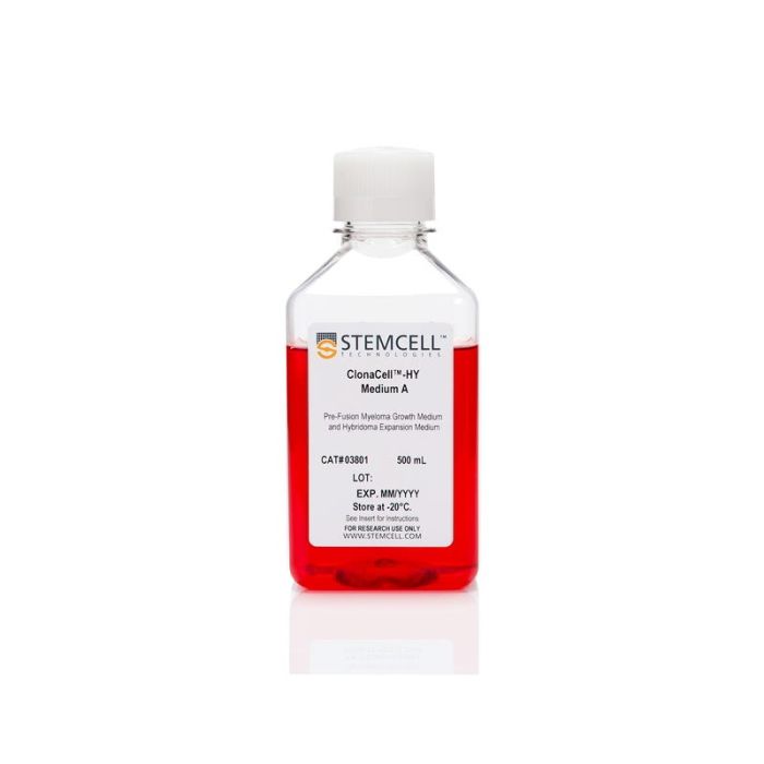
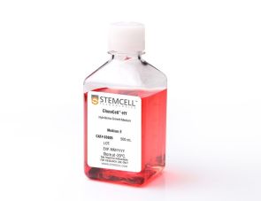
ClonaCell™-HY培养基A

骨髓瘤与杂交瘤培养基(含血清)
只有 %1
¥2,534.00

产品号 #(选择产品)

产品号 #03801


骨髓瘤与杂交瘤培养基(含血清)

总览

ClonaCell™-HY培养基A是一种含血清液体培养基,配方专为扩增融合前的亲本骨髓瘤细胞以及培养筛选后的稳定杂交瘤克隆而设计。该培养基经验证适用于小鼠和大鼠杂交瘤开发,据报告可用于生产人、小鼠、大鼠和仓鼠等多种宿主动物的淋巴细胞产生的杂交瘤。

包含
• DMEM培养基
• 血清
• 庆大霉素
• 2-巯基乙醇
• 酚红
• L-谷氨酰胺及其他添加物
• 其他成分
 
分类
专用培养基
 
细胞类型
杂交瘤细胞
 
种属
小鼠
 
应用
细胞培养,杂交瘤制备
 
品牌
ClonaCell
 
研究领域
抗体​制备,细胞系制备,杂交瘤制备
 

产品说明书及文档

请在《产品说明书》中查找相关支持信息和使用说明,或浏览下方更多实验方案。

Document Type
Product Name
Catalog #
Lot #
Language
Catalog #
03801
Lot #
All
Language
English
Document Type
Technical Manual
Catalog #
03801
Lot #
All
Language
English
Document Type
Safety Data Sheet
Catalog #
03801
Lot #
All
Language
English

应用领域

本产品专为以下研究领域设计,适用于工作流程中的高亮阶段。探索这些工作流程,了解更多我们为各研究领域提供的其他配套产品。

相关材料与文献

技术资料 (7)

常见问题

Why is there HT (hypoxanthine, thymidine) in Medium E?

Hybridomas are selected using HAT (hypoxanthine, aminopterin, thymidine). Aminopterin blocks the de novo pathway for synthesizing nucleotide precursors for DNA synthesis. The inhibition of the de novo pathway can persist even after the cells are removed from selection. Hypoxanthine and thymidine (HT) provide the necessary nucleotide precursors for hybridoma cells to synthesize DNA using the salvage pathway. Once the cells are growing well in Medium E, they can be gradually switched to Medium A or another medium without HT.

Is the serum in ClonaCell™-HY media heat inactivated?

Yes, all serum used in ClonaCell™-HY media is heat inactivated.

Is there any IgG in clonacell™-HY media?

While we don't add IgG to the ClonaCell™-HY media, we do add serum, which contains an undefined amount of IgG. We selectively use serum lots with low IgG levels in the production of ClonaCell™-HY media, however, levels vary from lot to lot. IgG levels in a specific lot of ClonaCell™-HY medium are available in the lot-specific Certificate of Analysis.

Are there antibiotics in ClonaCell™-HY media?

These products contain gentamycin rather than penicillin/streptomycin/amphotericin B, because gentamycin is more stable and is a broad spectrum antibiotic that is non-toxic to most mammalian cells in culture.

What is the optimal number of colonies per plate?

We recommend 50-150 colonies per plate. An average fusion will result in approximately 1000 colonies per fusion (approx. 100 colonies per plate). Even if the average number of colonies per plate approaches 300, there should still be enough separation between colonies to pick easily.

Why do I have to put my fused cells into liquid medium overnight? Why can't I just plate directly into Medium D?

We recommend waiting up to 24 hours so that all of the fused cells can go through one cell cycle. This will ensure they have a chance to express HPRT (hypoxanthine guanine phosphoribosyltransferase), the enzyme necessary to survive in the presence of aminopterin (present in Medium D). Additionally, fused cells are very fragile immediately after fusion. Waiting a day before mixing the cells with the methylcellulose will improve their survival. Although it is not recommended, fused cells may be plated on the same day as fusion, but the cells should be allowed to recover for several hours in ClonaCell™-HY Medium C prior to plating.

What myeloma and mouse strains should I use?

Myeloma: There are at least two common myeloma cell lines used to generate hybridomas - SP2/0 and P3X63Ag8.683. Both are available from ATCC. Researchers should ensure that the myeloma line is from a reliable source and is negative for mycoplasma. Mycoplasma contamination of the myeloma line can result in decreased efficiency of hybridoma formation. Mouse: We suggest using BALB/c splenocytes and parental myeloma cells of BALB/c for the following reasons: they are highly immune reactive, well characterized and myeloma cells are available from the same genetic strain. Other mouse strains, however, are also compatible with cloning in ClonaCell™-HY media.

Can I grow human/rat/T cell hybridomas in ClonaCell™-HY?

Although we have not tried to generate human, rat or T cell hybridomas during in-house testing, these experiments are expected to be successful using ClonaCell™-HY. The researcher would need to ensure that the cell lines used in the fusion are sensitive to HAT selection and grow well in methylcellulose-based medium.

There are very few colonies growing in my Medium D. Why?

Low numbers of colonies is generally a result of low fusion efficiency, which can have many causes. The fusion efficiency can be affected by the presence of serum during fusion, the presence of mycoplasma, low viability of cells, overexposure to polyethylene glycol or slow-growing myeloma cells prior to fusion.

Why does the ClonaCell™-HY manual suggest two different methods for fusion (A or B)? Can one expect better results with one method over the other?

Which method chosen is a personal preference and there should not be significant differences in efficiency. Method B is faster and has less steps, but Method B requires you to remove all the PEG before the cells are diluted, so you will risk aspirating cells if not very careful. With Method A, you dilute the PEG with Medium B, so you have less opportunity to lose cells.

Why does the ClonaCell™-HY manual suggest two different methods for fusion (A or B)? Can one expect better results with one method over the other?

A: Which method chosen is a personal preference and there should not be significant differences in efficiency. Method B is faster and has less steps, but Method B requires you to remove all the PEG before the cells are diluted, so you will risk aspirating cells if not very careful. With Method A, you dilute the PEG with Medium B, so you have less opportunity to lose cells.

Once I pick the colonies and grow the cells in plates, will the residual methylcellulose interfere with characterization? For example, will I have problems doing an ELISA?

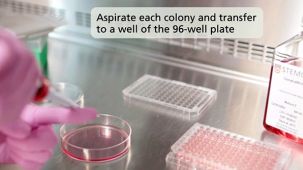
 There will likely be some residual methylcellulose contamination when colonies are picked and transferred to the 96-well plate with the liquid growth medium. The concentration of methylcellulose, however, should be low enough that it should not interfere with most assays.

Is the serum in ClonaCell™-TCS medium heat inactivated?

Yes, all serum used in ClonaCell™ is heat inactivated.

Is there any IgG in ClonaCell™ TCS?

While we don't add IgG to the ClonaCell™ media, we do add serum, which contains an undefined amount of IgG. We selectively use serum lots with low IgG levels in the production of ClonaCell™ media, however, levels vary from lot to lot. IgG levels in a specific lot of ClonaCell™ TCS medium are available in the lot-specific Certificate of Analysis.

Can ClonaCell™-TCS be used with any cell line?

A list of recommended cell lines can be found in the manual. Other cell lines may be compatible with ClonaCell™-TCS. It will be necessary, however, to determine the plating cell density and growth efficiency of the desired cells in ClonaCell™-TCS.

文献 (15)

Differential EphA2 epitope display on normal versus malignant cells. Coffman KT et al. Cancer Research 2003 NOV

Abstract

The EphA2 receptor tyrosine kinase is overexpressed in many different types of human cancers where it functions as a powerful oncoprotein. Dramatic changes in the subcellular localization and function of EphA2 have also been linked with cancer,and in particular,unstable cancer cell-cell contacts prevent EphA2 from stably binding its ligand on the surface of adjoining cells. This change is important in light of evidence that ligand binding causes EphA2 to transmit signals that negatively regulate tumor cell growth and invasiveness and also induce EphA2 degradation. On the basis of these properties,we have begun to target EphA2 on tumor cells using agonistic antibodies,which mimic the consequences of ligand binding. In our present study,we show that a subset of agonistic EphA2 antibodies selectively bind epitopes on malignant cells,which are not available on nontransformed epithelial cells. We also show that such epitopes arise from differential cell-cell adhesions and that the stable intercellular junctions of nontransformed epithelial cells occlude the binding site for ligand,as well as this subset of EphA2 antibodies. Finally,we demonstrate that antibody targeting of EphA2 decreases tumor cell growth as measured using xenograft tumor models and found that the mechanism of antibody action relates to EphA2 protein degradation in vivo. Taken together,these results suggest new opportunities for therapeutic targeting of the large number of different cancers that express EphA2 in a manner that could minimize potential toxicities to normal cells.
Carbohydrate mimicry between human ganglioside GM1 and Campylobacter jejuni lipooligosaccharide causes Guillain-Barre syndrome Yuki N et al. Proceedings of the National Academy of Sciences 2004 AUG

Abstract

Molecular mimicry between microbial and self-components is postulated as the mechanism that accounts for the antigen and tissue specificity of immune responses in postinfectious autoimmune diseases. Little direct evidence exists,and research in this area has focused principally on T cell-mediated,antipeptide responses,rather than on humoral responses to carbohydrate structures. Guillain-Barré syndrome,the most frequent cause of acute neuromuscular paralysis,occurs 1-2 wk after various infections,in particular,Campylobacter jejuni enteritis. Carbohydrate mimicry [Galbeta1-3GalNAcbeta1-4(NeuAcalpha2-3)Galbeta1-] between the bacterial lipooligosaccharide and human GM1 ganglioside is seen as having relevance to the pathogenesis of Guillain-Barré syndrome,and conclusive evidence is reported here. On sensitization with C. jejuni lipooligosaccharide,rabbits developed anti-GM1 IgG antibody and flaccid limb weakness. Paralyzed rabbits had pathological changes in their peripheral nerves identical with those present in Guillain-Barré syndrome. Immunization of mice with the lipooligosaccharide generated a mAb that reacted with GM1 and bound to human peripheral nerves. The mAb and anti-GM1 IgG from patients with Guillain-Barré syndrome did not induce paralysis but blocked muscle action potentials in a muscle-spinal cord coculture,indicating that anti-GM1 antibody can cause muscle weakness. These findings show that carbohydrate mimicry is an important cause of autoimmune neuropathy.
Generation of PRL-3- and PRL-1-specific monoclonal antibodies as potential diagnostic markers for cancer metastases Li J et al. Clinical Cancer Research 2005 MAR

Abstract

PURPOSE: The PRL-3 mRNA is consistently elevated in metastatic samples derived from colorectal cancers. We sought to generate a specific PRL-3 monoclonal antibody (mAb) that might serve as a potential diagnostic marker for colorectal cancer metastasis. EXPERIMENTAL DESIGN: PRL-3 is one of three members (PRL-1,PRL-2,and PRL-3) in a unique protein-tyrosine phosphatase family. Because the three PRLs are 76% to 87% identical in their amino acid sequences,it poses a great challenge to obtain mAbs that are specific for respective phosphatase of regenerating liver (PRL) but not for the other two in the family. We screened over 1,400 hybridoma clones to generate mAbs specific to each PRL member. RESULTS: We obtained two hybridoma clones specifically against PRL-3 and another two clones specifically against PRL-1. These antibodies had been evaluated by several critical tests to show their own specificities and applications. Most importantly,the PRL-3 mAbs were assessed on 282 human colorectal tissue samples (121 normal,17 adenomas,and 144 adenocarcinomas). PRL-3 protein was detected in 11% of adenocarcinoma samples. The PRL-3- and PRL-1-specific mAbs were further examined on 204 human multiple cancer tissues. The differential expressions of PRL-3 and PRL-1 confirmed the mAbs' specificity. CONCLUSIONS: Using several approaches,we show that PRL-3- or PRL-1-specific mAbs react only to their respective antigen. The expression of PRL-3 in textgreater10% of primary colorectal cancer samples indicates that PRL-3 may prime the metastatic process. These mAbs will be useful as markers in clinical diagnosis for assessing tumor aggressiveness.

更多信息

更多信息
物种 小鼠
Contains • DMEM • Serum • Gentamicin • 2-Mercaptoethanol • Phenol red • L-Glutamine and other supplements • Other ingredients
质量保证:

产品仅供研究使用,不用于针对人或动物的诊断或治疗。 欲获悉更多关于STEMCELL的质控信息,请访问 STEMCELL.CN/COMPLIANCE.
Copyright © 2026 by STEMCELL Technologies. All rights reserved.

在线联系