若您需要咨询产品或有任何技术问题,请通过官方电话 400 885 9050 或邮箱 info.cn@stemcell.com 与我们联系。

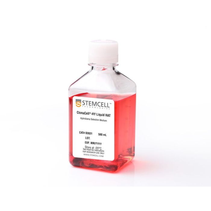
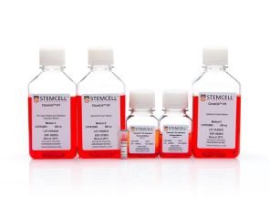
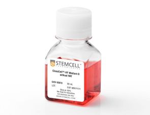
ClonaCell™-HY 液体 HAT 筛选培养基

用于筛选和克隆悬浮适应性细胞和贴壁细胞的半固体甲基纤维素基培养基(含血清)
只有 %1
¥5,228.00

产品号 #(选择产品)

产品号 #03831_C

用于筛选和克隆悬浮适应性细胞和贴壁细胞的半固体甲基纤维素基培养基(含血清)

产品优势

  • 简化液体悬浮培养中的杂交瘤筛选流程

总览

ClonaCell™-HY 液体 HAT 筛选培养基是一种含血清的液体培养基,含有次黄嘌呤、氨基蝶呤和胸苷(HAT),用于在淋巴细胞与骨髓瘤细胞融合后对杂交瘤进行筛选。该培养基已通过验证,可用于小鼠和大鼠杂交瘤的开发。据报道,该培养基也兼容多种宿主来源的淋巴细胞(包括人、小鼠、大鼠和仓鼠)在杂交瘤或骨髓瘤细胞的生产和克隆中的应用。

包含
• DMEM培养基
• 血清
• 次黄嘌呤、氨基蝶呤和胸苷 (HAT)
• 庆大霉素
• 2-巯基乙醇
• 酚红
• L-谷氨酰胺及其他添加物
• 其他成分
 
分类
专用培养基
 
细胞类型
杂交瘤细胞
 
种属
小鼠
 
应用
细胞培养,杂交瘤制备
 
品牌
ClonaCell
 
研究领域
抗体​制备,细胞系制备,杂交瘤制备
 

产品说明书及文档

请在《产品说明书》中查找相关支持信息和使用说明,或浏览下方更多实验方案。

Document Type
Product Name
Catalog #
Lot #
Language
Catalog #
03831
Lot #
All
Language
English
Document Type
Technical Manual
Catalog #
03831
Lot #
All
Language
English
Document Type
Safety Data Sheet
Catalog #
03831
Lot #
All
Language
English

应用领域

本产品专为以下研究领域设计,适用于工作流程中的高亮阶段。探索这些工作流程,了解更多我们为各研究领域提供的其他配套产品。

相关材料与文献

技术资料 (3)

常见问题

Is the serum in ClonaCell™-TCS medium heat inactivated?

Yes, all serum used in ClonaCell™ is heat inactivated.

Is there any IgG in ClonaCell™ TCS?

While we don't add IgG to the ClonaCell™ media, we do add serum, which contains an undefined amount of IgG. We selectively use serum lots with low IgG levels in the production of ClonaCell™ media, however, levels vary from lot to lot. IgG levels in a specific lot of ClonaCell™ TCS medium are available in the lot-specific Certificate of Analysis.

Can ClonaCell™-TCS be used with any cell line?

A list of recommended cell lines can be found in the manual. Other cell lines may be compatible with ClonaCell™-TCS. It will be necessary, however, to determine the plating cell density and growth efficiency of the desired cells in ClonaCell™-TCS.

Why is there HT (hypoxanthine, thymidine) in Medium E?

Hybridomas are selected using HAT (hypoxanthine, aminopterin, thymidine). Aminopterin blocks the de novo pathway for synthesizing nucleotide precursors for DNA synthesis. The inhibition of the de novo pathway can persist even after the cells are removed from selection. Hypoxanthine and thymidine (HT) provide the necessary nucleotide precursors for hybridoma cells to synthesize DNA using the salvage pathway. Once the cells are growing well in Medium E, they can be gradually switched to Medium A or another medium without HT.

Is the serum in ClonaCell™-HY media heat inactivated?

Yes, all serum used in ClonaCell™-HY media is heat inactivated.

Is there any IgG in clonacell™-HY media?

While we don't add IgG to the ClonaCell™-HY media, we do add serum, which contains an undefined amount of IgG. We selectively use serum lots with low IgG levels in the production of ClonaCell™-HY media, however, levels vary from lot to lot. IgG levels in a specific lot of ClonaCell™-HY medium are available in the lot-specific Certificate of Analysis.

Are there antibiotics in ClonaCell™-HY media?

These products contain gentamycin rather than penicillin/streptomycin/amphotericin B, because gentamycin is more stable and is a broad spectrum antibiotic that is non-toxic to most mammalian cells in culture.

What is the optimal number of colonies per plate?

We recommend 50-150 colonies per plate. An average fusion will result in approximately 1000 colonies per fusion (approx. 100 colonies per plate). Even if the average number of colonies per plate approaches 300, there should still be enough separation between colonies to pick easily.

Why do I have to put my fused cells into liquid medium overnight? Why can't I just plate directly into Medium D?

We recommend waiting up to 24 hours so that all of the fused cells can go through one cell cycle. This will ensure they have a chance to express HPRT (hypoxanthine guanine phosphoribosyltransferase), the enzyme necessary to survive in the presence of aminopterin (present in Medium D). Additionally, fused cells are very fragile immediately after fusion. Waiting a day before mixing the cells with the methylcellulose will improve their survival. Although it is not recommended, fused cells may be plated on the same day as fusion, but the cells should be allowed to recover for several hours in ClonaCell™-HY Medium C prior to plating.

What myeloma and mouse strains should I use?

Myeloma: There are at least two common myeloma cell lines used to generate hybridomas - SP2/0 and P3X63Ag8.683. Both are available from ATCC. Researchers should ensure that the myeloma line is from a reliable source and is negative for mycoplasma. Mycoplasma contamination of the myeloma line can result in decreased efficiency of hybridoma formation. Mouse: We suggest using BALB/c splenocytes and parental myeloma cells of BALB/c for the following reasons: they are highly immune reactive, well characterized and myeloma cells are available from the same genetic strain. Other mouse strains, however, are also compatible with cloning in ClonaCell™-HY media.

Can I grow human/rat/T cell hybridomas in ClonaCell™-HY?

Although we have not tried to generate human, rat or T cell hybridomas during in-house testing, these experiments are expected to be successful using ClonaCell™-HY. The researcher would need to ensure that the cell lines used in the fusion are sensitive to HAT selection and grow well in methylcellulose-based medium.

There are very few colonies growing in my Medium D. Why?

Low numbers of colonies is generally a result of low fusion efficiency, which can have many causes. The fusion efficiency can be affected by the presence of serum during fusion, the presence of mycoplasma, low viability of cells, overexposure to polyethylene glycol or slow-growing myeloma cells prior to fusion.

Why does the ClonaCell™-HY manual suggest two different methods for fusion (A or B)? Can one expect better results with one method over the other?

Which method chosen is a personal preference and there should not be significant differences in efficiency. Method B is faster and has less steps, but Method B requires you to remove all the PEG before the cells are diluted, so you will risk aspirating cells if not very careful. With Method A, you dilute the PEG with Medium B, so you have less opportunity to lose cells.

Why does the ClonaCell™-HY manual suggest two different methods for fusion (A or B)? Can one expect better results with one method over the other?

A: Which method chosen is a personal preference and there should not be significant differences in efficiency. Method B is faster and has less steps, but Method B requires you to remove all the PEG before the cells are diluted, so you will risk aspirating cells if not very careful. With Method A, you dilute the PEG with Medium B, so you have less opportunity to lose cells.

Once I pick the colonies and grow the cells in plates, will the residual methylcellulose interfere with characterization? For example, will I have problems doing an ELISA?

 There will likely be some residual methylcellulose contamination when colonies are picked and transferred to the 96-well plate with the liquid growth medium. The concentration of methylcellulose, however, should be low enough that it should not interfere with most assays.

文献 (7)

Immunoglobulin G specifically binding plant N-glycans with high affinity could be generated in rabbits but not in mice. Jin C et al. Glycobiology 2006 APR

Abstract

Xylosylated and core alpha1,3-fucosylated N-glycans from plants are immunogenic,and they play a still obscure role in allergy and in the field of plant-made protein pharmaceuticals. We immunized mice to generate monoclonal antibodies (mAbs) binding plant N-glycans specifically via the epitope containing either the xylose or the core alpha1,3-fucose residue. Splenocytes expressing N-glycan-specific antibodies derived from C57BL/6 mice previously immunized with plant glycoproteins were preselected by cell sorting to generate hybridoma lines producing specific antibodies. However,we obtained only mAbs unable to distinguish fucosylated from xylosylated N-glycans and reactive even with the pentasaccharide core Man3GlcNAc2. In contrast,immunization of rabbits yielded polyclonal sera selectively reactive with either fucosylated or xylosylated N-glycans. Purification of these sera using glyco-modified neoglycoproteins coupled to a chromatography matrix provided polyclonal sera suitable for affinity determination. Surface plasmon resonance measurements using sensor chips with immobilized glyco-modified transferrins revealed dissociation constants of around 10(-9) M. This unexpectedly high affinity of IgG antibodies toward carbohydrate epitopes has repercussions on our conception of the binding strength and significance of antiglycan IgE antibodies in allergy.
Antibody targeting to acClass I MHC-peptide epitope promotes tumor cell death Wittman VP et al. The Journal of Immunology 2006 SEP

Abstract

Therapeutic mAbs that target tumor-associated Ags on the surface of malignant cells have proven to be an effective and specific option for the treatment of certain cancers. However,many of these protein markers of carcinogenesis are not expressed on the cells' surface. Instead these tumor-associated Ags are processed into peptides that are presented at the cell surface,in the context of MHC class I molecules,where they become targets for T cells. To tap this vast source of tumor Ags,we generated a murine IgG2a mAb,3.2G1,endowed with TCR-like binding specificity for peptide-HLA-A*0201 (HLA-A2) complex and designated this class of Ab as TCR mimics (TCRm). The 3.2G1 TCRm recognizes the GVL peptide (GVLPALPQV) from human chorionic gonadotropin beta presented by the peptide-HLA-A*0201 complex. When used in immunofluorescent staining reactions using GVL peptide-loaded T2 cells,the 3.2G1 TCRm specifically stained the cells in a peptide and Ab concentration-dependent manner. Staining intensity correlated with the extent of cell lysis by complement-dependent cytotoxicity (CDC),and a peptide concentration-dependent threshold level existed for the CDC reaction. Staining of human tumor lines demonstrated that 3.2G1 TCRm was able to recognize endogenously processed peptide and that the breast cancer cell line MDA-MB-231 highly expressed the target epitope. The 3.2G1 TCRm-mediated CDC and Ab-dependent cellular cytotoxicity of a human breast carcinoma line in vitro and inhibited in vivo tumor implantation and growth in nude mice. These results provide validation for the development of novel TCRm therapeutic reagents that specifically target and kill tumors via recognition and binding to MHC-peptide epitopes.
Levels of specific peptide-HLA class I complex predicts tumor cell susceptibility to CTL killing. Weidanz Ja et al. Journal of Immunology (Baltimore,Md. : 1950) 2006 OCT

Abstract

Recognition of tumor-associated Ags (TAAs) on tumor cells by CTLs and the subsequent tumor cell death are assumed to be dependent on TAA protein expression and to correlate directly with the level of peptide displayed in the binding site of the HLA class I molecule. In this study we evaluated whether the levels of Her-2/neu protein expression on human tumor cell lines directly correlate with HLA-A*0201/Her2/neu peptide presentation and CTL recognition. We developed a TCR mimic (TCRm) mAb designated 1B8 that specifically recognizes the HLA-A2.1/Her2/neu peptide (369-377) (Her2(369)-A2) complex. TCRm mAb staining intensity varied for the five human tumor cell lines analyzed,suggesting quantitative differences in levels of the Her2(369)-A2 complex on these cells. Analysis of tumor cell lines pretreated with IFN-gamma and TNF-alpha for Her2/neu protein and HLA-A2 molecule expression did not reveal a direct correlation between the levels of Her2/neu Ag,HLA-A2 molecule,and Her2(369)-A2 complex expression. However,compared with untreated cells,cytokine-treated cell lines showed an increase in Her2(369)-A2 epitope density that directly correlated with enhanced tumor cell death (p = 0.05). Although a trend was observed between tumor cell lysis and the level of the Her2(369)-A2 complex for untreated cells,the association was not significant. These findings suggest that tumor cell susceptibility to CTL-mediated lysis may be predicted based on the level of specific peptide-MHC class I expression rather than on the total level of TAA expression. Further,these studies demonstrate the potential of the TCRm mAb for validation of endogenous HLA-peptide epitopes on tumor cells.

更多信息

更多信息
物种 小鼠
Contains • DMEM • Serum • Hypoxanthine, aminopterin, and thymidine (HAT) • Gentamicin • 2-Mercaptoethanol • Phenol red • L-Glutamine and other supplements • Other ingredients
质量保证:

产品仅供研究使用,不用于针对人或动物的诊断或治疗。 欲获悉更多关于STEMCELL的质控信息,请访问 STEMCELL.CN/COMPLIANCE. Safety Statement: CA WARNING: This product can expose you to Aminopterin which is known to the State of California to cause birth defects or other reproductive harm. For more information go to www.P65Warnings.ca.gov
Copyright © 2026 by STEMCELL Technologies. All rights reserved.

在线联系