若您需要咨询产品或有任何技术问题,请通过官方电话 400 885 9050 或邮箱 info.cn@stemcell.com 与我们联系。

ClonaCell™-HY杂交瘤试剂盒

杂交瘤制备全流程解决方案
只有 %1
¥19,268.00

产品号 #(选择产品)

产品号 #03800


杂交瘤制备全流程解决方案

产品优势

  • 省时优势:将杂交瘤筛选与克隆整合为单步操作
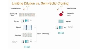
  • 资源节约:单细胞来源的杂交瘤在半固体培养基中形成可见的离散集落,便于挑取并转移至液体培养基进行筛查和扩增
  • 高克隆效率:单个细胞被悬浮固定于半固体培养基中,避免高产出的稀有克隆因竞争性生长而丢失

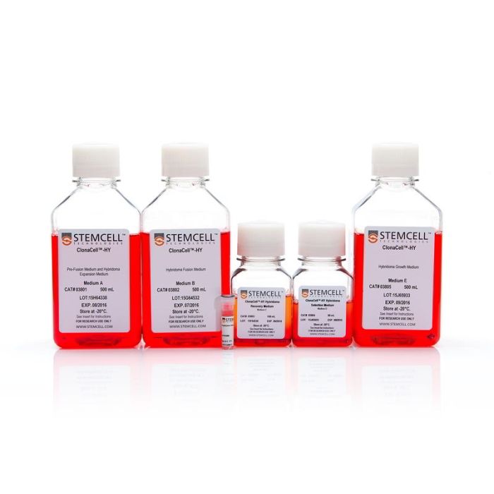
产品组分包括

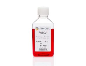
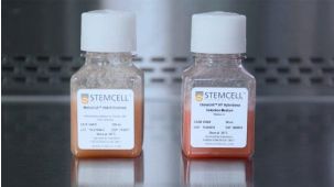
  • ClonaCell™-HY培养基A,500 mL(产品号 #03801)
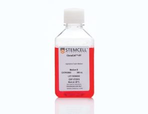
  • ClonaCell™-HY培养基B,500 mL(产品号 #03802)
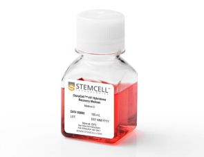
  • ClonaCell™-HY培养基C,100 mL(产品号 #03803)
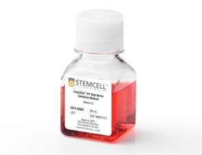
  • ClonaCell™-HY培养基D,90 mL(产品号 #03804)
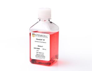
  • ClonaCell™-HY培养基E,500 mL(产品号 #03805)
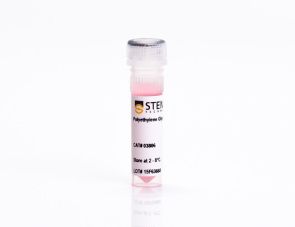
  • ClonaCell™-HY PEG,1.5 mL(产品号 #03806)

总览

使用ClonaCell™-HY杂交瘤试剂盒中包含的培养基和试剂,完成杂交瘤开发及单克隆抗体生产的所有步骤:
ClonaCell™-HY培养基A用于骨髓瘤和杂交瘤培养
ClonaCell™-HY培养基B用于杂交瘤融合
ClonaCell™-HY培养基C用于杂交瘤融合后恢复
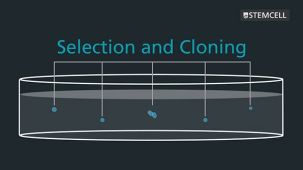
ClonaCell™-HY培养基D用于杂交瘤筛选与克隆
ClonaCell™-HY培养基E用于杂交瘤生长
ClonaCell™-HY PEG用于支持杂交瘤融合

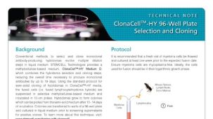
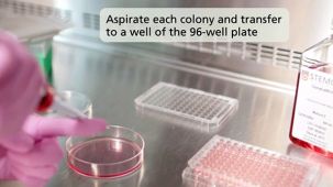
ClonaCell™-HY方法采用基于甲基纤维素的半固体选择性培养基,将杂交瘤筛选和克隆合二为一。在半固体培养基中,单个亲代杂交瘤克隆及其子代在生长形成独立克隆的过程中保持聚集状态。这避免了因生长较快的细胞过度生长而导致稀有克隆丢失的情况,这种情况可能发生在液体培养基筛选过程中。杂交瘤克隆可通过人工或仪器轻松地从半固体培养基中挑取,并分散到液体培养基中进行筛选和扩增。

该试剂盒已验证可用于小鼠和大鼠杂交瘤的构建和单克隆抗体的生产,并兼容多种宿主物种(包括人、小鼠、大鼠和仓鼠)的淋巴细胞,可用于杂交瘤的生产、克隆和扩增。为方便起见,该试剂盒各组分也可单独购买。


分类
半固体培养基,专用培养基
 
细胞类型
杂交瘤细胞
 
种属
小鼠,其他物种,大鼠
 
应用
细胞培养,杂交瘤制备
 
品牌
ClonaCell
 
研究领域
抗体​制备,细胞系制备,杂交瘤制备
 

产品说明书及文档

请在《产品说明书》中查找相关支持信息和使用说明,或浏览下方更多实验方案。

Document Type
Product Name
Catalog #
Lot #
Language
Catalog #
03800
Lot #
All
Language
English
Catalog #
03800
Lot #
All
Language
English
Catalog #
03800
Lot #
All
Language
English
Catalog #
03800
Lot #
All
Language
English
Document Type
Technical Manual
Catalog #
03800
Lot #
All
Language
English
Document Type
Safety Data Sheet
Catalog #
03800
Lot #
All
Language
English

应用领域

本产品专为以下研究领域设计,适用于工作流程中的高亮阶段。探索这些工作流程,了解更多我们为各研究领域提供的其他配套产品。

相关材料与文献

技术资料 (11)

常见问题 (12)

Why is there HT (hypoxanthine, thymidine) in Medium E?

Hybridomas are selected using HAT (hypoxanthine, aminopterin, thymidine). Aminopterin blocks the de novo pathway for synthesizing nucleotide precursors for DNA synthesis. The inhibition of the de novo pathway can persist even after the cells are removed from selection. Hypoxanthine and thymidine (HT) provide the necessary nucleotide precursors for hybridoma cells to synthesize DNA using the salvage pathway. Once the cells are growing well in Medium E, they can be gradually switched to Medium A or another medium without HT.

Is the serum in ClonaCell™-HY media heat inactivated?

Yes, all serum used in ClonaCell™-HY media is heat inactivated.

Is there any IgG in clonacell™-HY media?

While we don't add IgG to the ClonaCell™-HY media, we do add serum, which contains an undefined amount of IgG. We selectively use serum lots with low IgG levels in the production of ClonaCell™-HY media, however, levels vary from lot to lot. IgG levels in a specific lot of ClonaCell™-HY medium are available in the lot-specific Certificate of Analysis.

Are there antibiotics in ClonaCell™-HY media?

These products contain gentamycin rather than penicillin/streptomycin/amphotericin B, because gentamycin is more stable and is a broad spectrum antibiotic that is non-toxic to most mammalian cells in culture.

What is the optimal number of colonies per plate?

We recommend 50-150 colonies per plate. An average fusion will result in approximately 1000 colonies per fusion (approx. 100 colonies per plate). Even if the average number of colonies per plate approaches 300, there should still be enough separation between colonies to pick easily.

Why do I have to put my fused cells into liquid medium overnight? Why can't I just plate directly into Medium D?

We recommend waiting up to 24 hours so that all of the fused cells can go through one cell cycle. This will ensure they have a chance to express HPRT (hypoxanthine guanine phosphoribosyltransferase), the enzyme necessary to survive in the presence of aminopterin (present in Medium D). Additionally, fused cells are very fragile immediately after fusion. Waiting a day before mixing the cells with the methylcellulose will improve their survival. Although it is not recommended, fused cells may be plated on the same day as fusion, but the cells should be allowed to recover for several hours in ClonaCell™-HY Medium C prior to plating.

What myeloma and mouse strains should I use?

Myeloma: There are at least two common myeloma cell lines used to generate hybridomas - SP2/0 and P3X63Ag8.683. Both are available from ATCC. Researchers should ensure that the myeloma line is from a reliable source and is negative for mycoplasma. Mycoplasma contamination of the myeloma line can result in decreased efficiency of hybridoma formation. Mouse: We suggest using BALB/c splenocytes and parental myeloma cells of BALB/c for the following reasons: they are highly immune reactive, well characterized and myeloma cells are available from the same genetic strain. Other mouse strains, however, are also compatible with cloning in ClonaCell™-HY media.

Can I grow human/rat/T cell hybridomas in ClonaCell™-HY?

Although we have not tried to generate human, rat or T cell hybridomas during in-house testing, these experiments are expected to be successful using ClonaCell™-HY. The researcher would need to ensure that the cell lines used in the fusion are sensitive to HAT selection and grow well in methylcellulose-based medium.

There are very few colonies growing in my Medium D. Why?

Low numbers of colonies is generally a result of low fusion efficiency, which can have many causes. The fusion efficiency can be affected by the presence of serum during fusion, the presence of mycoplasma, low viability of cells, overexposure to polyethylene glycol or slow-growing myeloma cells prior to fusion.

Why does the ClonaCell™-HY manual suggest two different methods for fusion (A or B)? Can one expect better results with one method over the other?

Which method chosen is a personal preference and there should not be significant differences in efficiency. Method B is faster and has less steps, but Method B requires you to remove all the PEG before the cells are diluted, so you will risk aspirating cells if not very careful. With Method A, you dilute the PEG with Medium B, so you have less opportunity to lose cells.

Why does the ClonaCell™-HY manual suggest two different methods for fusion (A or B)? Can one expect better results with one method over the other?

A: Which method chosen is a personal preference and there should not be significant differences in efficiency. Method B is faster and has less steps, but Method B requires you to remove all the PEG before the cells are diluted, so you will risk aspirating cells if not very careful. With Method A, you dilute the PEG with Medium B, so you have less opportunity to lose cells.

Once I pick the colonies and grow the cells in plates, will the residual methylcellulose interfere with characterization? For example, will I have problems doing an ELISA?

 There will likely be some residual methylcellulose contamination when colonies are picked and transferred to the 96-well plate with the liquid growth medium. The concentration of methylcellulose, however, should be low enough that it should not interfere with most assays.

文献 (71)

Depletion of eosinophils in mice through the use of antibodies specific for C-C chemokine receptor 3 (CCR3). Grimaldi JC et al. Journal of Leukocyte Biology 1999 JUN

Abstract

We have generated rat monoclonal antibodies specific for the mouse eotaxin receptor,C-C chemokine receptor 3 (CCR3). Several anti-CCR3 mAbs proved to be useful for in vivo depletion of CCR3-expressing cells and immunofluorescent staining. In vivo CCR3 mAbs of the IgG2b isotype substantially depleted blood eosinophil levels in Nippostrongyus brasiliensis-infected mice. Repeated anti-CCR3 mAb treatment in these mice significantly reduced tissue eosinophilia in the lung tissue and bronchoalveolar lavage fluid. Flow cytometry revealed that mCCR3 was expressed on eosinophils but not on stem cells,dendritic cells,or cells from the thymus,lymph node,or spleen of normal mice. Unlike human Th2 cells,mouse Th2 cells did not express detectable levels of CCR3 nor did they give a measurable response to eotaxin. None of the mAbs were antagonists or agonists of CCR3 calcium mobilization. To our knowledge,the antibodies described here are the first mAbs reported to be specific for mouse eosinophils and to be readily applicable for the detection,isolation,and in vivo depletion of eosinophils.
Protein phosphatase 2Calpha dephosphorylates axin and activates LEF-1-dependent transcription. Strovel ET et al. The Journal of biological chemistry 2000 JAN

Abstract

The Dishevelled (Dvl) gene family encodes cytoplasmic proteins that are necessary for Wnt signal transduction. Utilizing the yeast two-hybrid system,we identified protein phosphatase 2Calpha (PP2C) as a Dvl-PDZ domain-interacting protein. PP2C exists in a complex with Dvl,beta-catenin,and Axin,a negative regulator of Wnt signaling. In a Wnt-responsive LEF-1 reporter gene assay,expression of PP2C activates transcription and also elicits a synergistic response with beta-catenin and Wnt-1. In addition,PP2C expression relieves Axin-mediated repression of LEF-1-dependent transcription. PP2C utilizes Axin as a substrate both in vitro and in vivo and decreases its half-life. These results indicate that PP2C is a positive regulator of Wnt signal transduction and mediates its effects through the dephosphorylation of Axin.
Endogenous protein kinase CK2 participates in Wnt signaling in mammary epithelial cells Song DH et al. Journal of Biological Chemistry 2000 AUG

Abstract

Protein kinase CK2 (formerly casein kinase II) is a serine/threonine kinase overexpressed in many human tumors,transformed cell lines,and rapidly proliferating tissues. Recent data have shown that many cancers involve inappropriate reactivation of Wnt signaling through ectopic expression of Wnts themselves,as has been seen in a number of human breast cancers,or through mutation of intermediates in the Wnt pathway,such as adenomatous polyposis coli or beta-catenin,as described in colon and other cancers. Wnts are secreted factors that are important in embryonic development,but overexpression of certain Wnts,such as Wnt-1,leads to proliferation and transformation of cells. We report that upon stable transfection of Wnt-1 into the mouse mammary epithelial cell line C57MG,morphological changes and increased proliferation are accompanied by increased levels of CK2,as well as of beta-catenin. CK2 and beta-catenin co-precipitate with the Dvl proteins,which are Wnt signaling intermediates. A major phosphoprotein of the size of beta-catenin appears in in vitro kinase reactions performed on the Dvl immunoprecipitates. In vitro translated beta-catenin,Dvl-2,and Dvl-3 are phosphorylated by CK2. The selective CK2 inhibitor apigenin blocks proliferation of Wnt-1-transfected cells,abrogates phosphorylation of beta-catenin,and reduces beta-catenin and Dvl protein levels. These results demonstrate that endogenous CK2 is a positive regulator of Wnt signaling and growth of mammary epithelial cells.

更多信息

更多信息
物种 其它物种, 大鼠, 小鼠
质量保证:

产品仅供研究使用,不用于针对人或动物的诊断或治疗。 欲获悉更多关于STEMCELL的质控信息,请访问 STEMCELL.CN/COMPLIANCE.
Copyright © 2026 by STEMCELL Technologies. All rights reserved.

在线联系