产品号 #05790_C
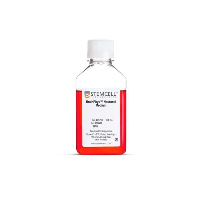
提升神经元功能的无血清基础培养基
若您需要咨询产品或有任何技术问题,请通过官方电话 400 885 9050 或邮箱 info.cn@stemcell.com 与我们联系。
I want to help neuroscientists like you create more physiological culture conditions, for more active and healthy neuronal cultures.

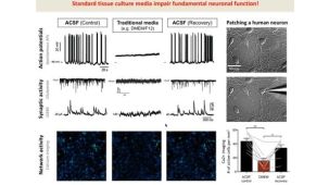
BrainPhys™神经元培养基以Bardy和Gage(Bardy et al, PNAS, 2015)的配方为基础,是一种无血清的基础培养基,可模拟中枢神经系统(CNS)细胞外环境,经过优化可促进而非抑制培养的原代或人多能干细胞(hPSC)衍生神经元的神经元活性和成熟,从而获得更高比例的具有突触活性的神经元。
BrainPhys™神经元培养基适用于hPSC和CNS衍生神经元的长期培养。为避免在更换培养基时对细胞造成冲击,您也可以在进行功能性实验(例如基于微电极阵列记录或活细胞荧光成像)时使用BrainPhys™培养基。
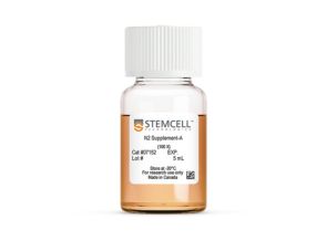
为确保细胞在长期无血清培养中的健康,BrainPhys™神经元培养基需搭配合适的血清替代添加物使用。对于培养hPSC衍生神经元的研究人员,BrainPhys™ hPSC神经元试剂盒提供可BrainPhys™神经元培养基以及推荐的添加物,从而简化hPSC的分化和成熟。对于原代神经元培养,BrainPhys™原代神经元试剂盒提供了完整的、针对CNS衍生神经元优化的解决方案。如果您想要定制您的工作流程,BrainPhys™神经元培养基也以其他试剂盒形式提供,如只包含单独的NeuroCult™ SM1神经元添加物(BrainPhys™神经元培养基和SM1试剂盒),或与N2添加物-A组合(BrainPhys™神经元培养基N2-A & SM1试剂盒)。
查看我们的其他资源,了解更多关于BrainPhys™产品线的信息。
分类
基础培养基,专用培养基
细胞类型
神经细胞,PSC衍生,神经元,多能干细胞
种属
人,小鼠,大鼠
应用
细胞培养,分化,培养
品牌
BrainPhys
研究领域
疾病建模,药物发现和毒理检测,神经科学,干细胞生物学
制剂类别
无血清
Table 1. Properties of Culture Media (C Bardy et al. Proc Natl Acad Sci USA, 2015)
Check-mark denotes physiological conditions and supported activities according to C Bardy et al. Proc Natl Acad Sci USA, 2015.
Figure 1. Protocol for Plating and Culturing Primary Neurons with the SM1 Culture System
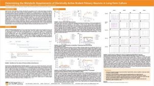
Primary rodent tissue dissociated in papain was plated in NeuroCult™ Neuronal Plating Medium, supplemented with NeuroCult™ SM1 Neuronal Supplement, L-Glutamine, and L-Glutamic Acid. On day 5, primary neurons were transitioned to BrainPhys™ Neuronal Medium, supplemented with NeuroCult™ SM1 Neuronal Supplement, by performing half-medium changes every 3 - 4 days.
Figure 2. Protocol for Culturing hPSCs with the SM1 Culture System
hPSCs were maintained in mTeSR™1 medium and then differentiated using the STEMdiff™ SMADi Neural Induction Kit. Following plating on PLO/laminin, half-medium changes were performed to transition to BrainPhys™ Neuronal Medium for maturation and long-term culture.
Figure 3. The SM1 Culture System Supports Long-Term Culture of Rodent Neurons
Primary E18 rat cortical neurons were cultured in the SM1 Culture System. A large number of viable neurons are visible after (A) 21 and (B) 35 days, as demonstrated by their bright neuronal cell bodies, and extensive neurite outgrowth and branching. Neurons are evenly distributed over the culture surface with minimal cell clumping.
Figure 4. Pre- and Post-Synaptic Markers are Expressed in Rodent Neurons Cultured in the SM1 Culture System
Primary E18 rat cortical neurons were cultured in the SM1 Culture System. At 21 DIV, neurons are phenotypically mature, as indicated by the presence of an extensive dendritic arbor, and appropriate expression and localization of pre-synaptic synapsin (A,C; green) and post-synaptic PSD-95 (A,B; red) markers. Synapsin is concentrated in discrete puncta distributed along the somata and dendritic processes, as defined by the dendritic marker MAP2 (A,D; blue).
Figure 5. The SM1 Culture System Supports Increased Cell Survival
(A) Primary E18 rat cortical neurons were cultured in the SM1 Culture System or a Competitor Culture System for 21 days. Neurons cultured in the SM1 Culture System have a significantly higher number of viable cells compared to the competitor culture system (n = 4; mean ± 95% CI; *p < 0.05). (B) Primary E18 rat cortical neurons were cultured in Neurobasal® supplemented with NeuroCult™ SM1 Neuronal Supplement (SM1) or competitor B27-like supplements (Competitor 1,2,3) for 21 days. Cultures supplemented with NeuroCult™ SM1 Neuronal Supplement have an equal number of neurons compared to competitor-supplemented cultures. Bars represent standard error of mean.
Figure 6. BrainPhys™ Supports Improved Neuronal Activity and More Consistent Network Bursting in Long-Term Culture
Raster plots from MEA recordings show the firing patterns of primary E18 rat cortical neurons across 8 electrodes at Weeks 2, 4, 6 and 8. Neurons were either cultured with a Commercial Medium with Supplements, Commercial Medium Plus with Supplements, BrainPhys™ and SM1, or BrainPhys™ and SM1 with 15 mM glucose. Detected spikes (black lines), single channel bursts (blue lines; a collection of at least 5 spikes, each separated by an ISI of no more than 100 ms), and network bursts (magenta boxes; a collection of at least 50 spikes from a minimum of 35% of participating electrodes across each well, each separated by an ISI of no more than 100 ms) were recorded for each medium. (A-D) Neurons cultured with Commercial Medium exhibited network bursting in Week 2 but no spiking activity was detected in subsequent timepoints. (E-H) In Commercial Medium Plus-cultured neurons, a high number of spikes and regular network bursting were detected at Week 2. A decreased number of spikes and inconsistent network bursting were observed in later time points, corresponding to the drop in MFR seen in Figure 4. (I-L) Without glucose, individual spiking was observed at Weeks 2 and 4 with BrainPhys™ and SM1 but network bursting was not detected until Weeks 6 and 8. (M-T) In contrast, neurons cultured with BrainPhys™ and SM1 with 15 mM glucose demonstrated strong spiking activity and consistent network bursting at all timepoints. MEA = microelectrode array; ISI = inter-spike interval; MFR = mean firing rate
Figure 7. Glucose Supplementation in BrainPhys™ Maintains Neuronal Activity Over 8 Weeks in Culture
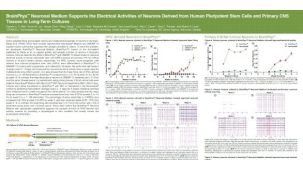
Primary E18 rat cortical neurons were cultured with BrainPhys™ and SM1 or other commercially available culture systems for 8 weeks. Neuronal activity can be detected at Day 9 with BrainPhys™, whereas activity is not detected until Day 14 in cultures maintained in either of the Commercial Media with Commercial Supplements. For Commercial Medium and Supplement-cultured neurons, mean firing rate remains low throughout culture. In contrast, a “peak-drop” activity pattern is observed in the Commercial Medium Plus condition, where mean firing rate increases rapidly within 2 days, followed by a drop in activity in the next 2 - 4 days. BrainPhys™and SM1 Kit with 15 mM glucose maintains the highest level of activity throughout the 8-week culture period.
Figure 8. hPSC-Derived Neurons Generated in BrainPhys™ Neuronal Medium Express Markers of Neuronal Maturity After 14 and 44 Days of Differentiation
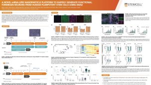
NPCs were generated from H9 cells using STEMdiff™ Neural Induction Medium in an embryoid body-based protocol. Next, NPCs were cultured in (A,C) BrainPhys™ Neuronal Medium, supplemented with 2% NeuroCult™ SM1 Supplement, 1% N2 Supplement-A, 20 ng/mL GDNF, 20 ng/mL BDNF, 1 mM db-cAMP and 200 nM ascorbic acid to initiate neuronal differentiation, or (B,D) DMEM/F12 under the same supplementation conditions. After 14 and 44 days of differentiation and maturation, neurons express the synaptic marker Synapsin 1 (green) and the mature neuronal marker MAP2 (red). In this example, neurons matured in BrainPhys™ Neuronal Medium show increased Synapsin 1 staining. Scale bar= 100 µm
Figure 9. hPSC-Derived Neurons Generated in BrainPhys™ Neuronal Medium and NeuroCult™ SM1 and N2 Supplements are Healthy and Morphologically Normal
NPCs were generated from H9 cells using STEMdiff™ Neural Induction Medium in an embryoid body-based protocol. Next, NPCs were cultured for 44 DIV in (A) BrainPhys™ Neuronal Medium, supplemented with 2% NeuroCult™ SM1 Supplement, 1% N2 Supplement-A, 20 ng/mL GDNF, 20 ng/mL BDNF, 1 mM db-cAMP and 200 nM ascorbic acid to initiate neuronal differentiation, or (B) DMEM/F12 under the same supplementation conditions. Neuronal cultures differentiated from NPCs in BrainPhys™ Neuronal Medium display extensive neurite outgrowth and reduced cellular debris compared to cultures differentiated in DMEM/F12. Scale bar= 100 µm.
Figure 10. hPSC-Derived Neurons Matured in BrainPhys™ Neuronal Medium Show Improved Excitatory and Inhibitory Synaptic Activity
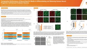
NPCs were generated from H9 cells using STEMdiff™ Neural Induction Medium in an embryoid body-based protocol. Next, NPCs were cultured for 44 DIV in (A,C) BrainPhys™ Neuronal Medium, supplemented with 2% NeuroCult™ SM1 Supplement, 1% N2 Supplement-A, 20 ng/mL GDNF, 20 ng/mL BDNF, 1 mM db-cAMP and 200 nM ascorbic acid to initiate neuronal differentiation, or (B,D) in DMEM/F12 under the same supplementation conditions. (A,C) Neurons matured in BrainPhys™ Neuronal Medium showed spontaneous excitatory (AMPA-mediated; A) and inhibitory (GABA-mediated; C) synaptic events. The frequency and amplitude of spontaneous synaptic events is consistently greater in neuronal cultures matured in BrainPhys™ Neuronal Medium, compared to neurons plated and matured in DMEM/F12 (B,D). Traces are representative.
请在《产品说明书》中查找相关支持信息和使用说明,或浏览下方更多实验方案。
本产品专为以下研究领域设计,适用于工作流程中的高亮阶段。探索这些工作流程,了解更多我们为各研究领域提供的其他配套产品。
Thank you for your interest in IntestiCult™ Organoid Growth Medium (Human). Please provide us with your contact information and your local representative will contact you with a customized quote. Where appropriate, they can also assist you with a(n):
Estimated delivery time for your area
Product sample or exclusive offer
In-lab demonstration
| 物种 | 人, 大鼠, 小鼠 |
|---|---|
| 配方 | 无血清 |
无血清神经添加物(50X)
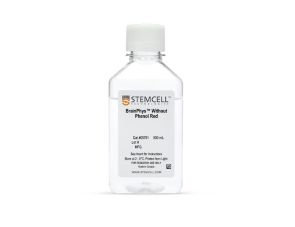
提升神经元功能的无血清基础培养基
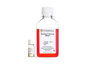
试剂盒包括BrainPhys™神经元培养基和SM1神经元添加物,用于原代神经元以及胚胎干/诱导多能干细胞来源神经元的无血清培养
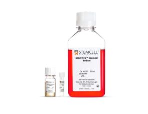
用于在BrainPhys™神经元培养基中无血清培养ES/iPS细胞来源的神经元
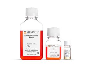
BrainPhys™ 原代神经元无血清培养试剂盒
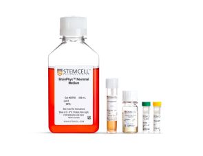
用于在BrainPhys™神经元培养基中无血清培养和分化ES/iPS细胞来源的神经元的试剂盒
无血清神经生理基础培养基,改善神经元活细胞成像和功能
用于小鼠和人胚胎干细胞和iPS细胞的神经和胰腺分化
扫描二维码或搜索微信号STEMCELLTech,即可关注我们的微信平台,第一时间接收丰富的技术资源和最新的活动信息。
如您有任何问题,欢迎发消息给STEMCELLTech微信公众平台,或与我们通过电话/邮件联系:400 885 9050 INFO.CN@STEMCELL.COM。
在线联系