若您需要咨询产品或有任何技术问题,请通过官方电话 400 885 9050 或邮箱 info.cn@stemcell.com 与我们联系。

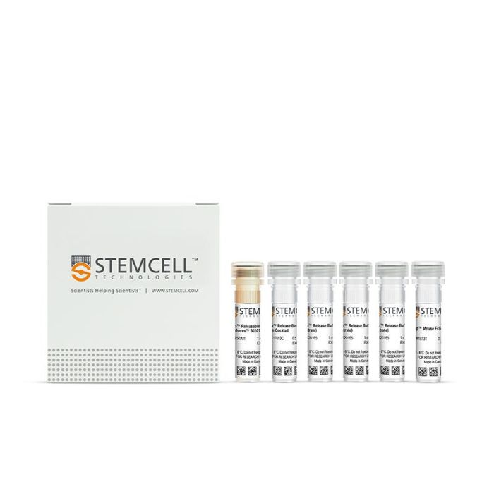
EasySep™ Release小鼠Biotin正选试剂盒

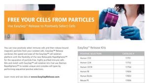
采用可解离磁珠技术对生物素偶联抗体标记的小鼠细胞进行免疫磁珠正选

只有 %1
¥11,648.00

产品号 #(选择产品)

产品号 #17655_C

通过免选磁珠正选分离出不含磁珠的生物素化抗体标记小鼠细胞

产品优势

  • 只需不到40分钟,从小鼠组织中分离出用生物素化抗体标记的高纯度细胞
  • 无需洗涤去除EasySep™ Releasable RapidSpheres™可解离磁珠

产品组分包括

  • EasySep™ Release小鼠Biotin正选试剂盒(产品号 #17655)
    • EasySep™Biotin正选抗体混合物,0.5 mL
    • EasySep™ Releasable RapidSpheres™ 50201磁珠,1 mL
    • EasySep™Release缓冲液(浓缩),3 x 1 mL
  • EasySep™小鼠FcR阻断剂(产品号 #18731),0.5 mL
专为您的实验方案打造的产品
要查看实验方案所需的所有配套产品,请参阅《实验方案与技术文档》

总览

使用 EasySep™ Release小鼠Biotin正选试剂盒,可通过免疫磁性正选,轻松从小鼠脾细胞中分离出高纯度、且不含磁珠的生物素化抗体标记的小鼠细胞。EasySep™技术结合单克隆抗体的特异性和无柱磁珠分选系统的简便性,已在发表的研究中广泛应用超过20年。

该EasySep™正选流程中,首先用能识别生物素和磁珠(称为EasySep™可解离磁珠)的抗体复合物标记目标细胞。与传统磁珠结合目标细胞不同,这些磁珠具有可解离的特性。经EasySep™磁极分离后,目的细胞首先被抗体和特异性磁珠标记,非目的细胞通过简单倾倒弃去,而目的细胞则保留在管中。随后,使用解离试剂将结合的磁珠从经EasySep™分选、带有生物素抗体标记的细胞上解离。仅需不到40分钟完成分选,目的细胞可立即用于流式细胞术、细胞培养或DNA/RNA提取等下游应用。使用该EasySep™试剂盒分选之后,细胞表面仍结合有抗体复合物,并可能与Brilliant Violet™偶联的抗体、聚乙二醇修饰的蛋白质或其他化学相关配体相互作用。 

了解更多关于免疫磁珠EasySep™技术的工作原理。探索更多为您的实验流程优化的产品,包括培养基、添加物、抗体等配套试剂。


磁极兼容性
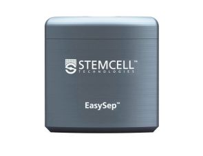
• EasySep™磁极(产品号 #18000)
• “The Big Easy” EasySep™磁极(产品号 #18001)
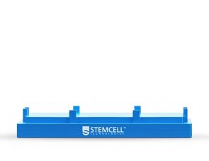
• EasyPlate™ EasySep™磁极(产品号 #18102)
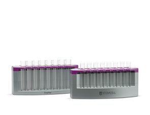
• EasyEights™ EasySep™磁极(产品号 #18103)
 
分类
细胞分选试剂盒
 
细胞类型
B 细胞,树突状细胞(DCs),粒细胞及其亚群,造血干/祖细胞,先天性淋巴细胞,淋巴细胞,巨噬细胞,骨髓基质细胞,间充质干/祖细胞,单核细胞,单个核细胞,髓系细胞,NK 细胞,其他组织,血浆,T 细胞
 
种属
小鼠
 
样本来源
骨髓,其他组织,脾脏
 
分选方法
正选
 
应用
细胞分选
 
品牌
EasySep
 
研究领域
免疫
 

实验数据

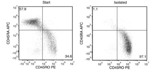
Typical EasySep™ Release Mouse Biotin Positive Selection Kit Cell Isolation Profile from PBMCs

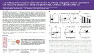
Figure 1. Typical EasySep™ Release Mouse Biotin Positive Selection Kit Cell Isolation Profile from PBMCs

Starting with fresh PBMCs, the purities of the start and final isolated fractions are 34.6% and 97.1%, respectively, using a biotinylated anti-human CD45RO antibody and EasySep™ Release Human Biotin Positive Selection Kit (as assessed by labeling with CD45RO and CD45RA).

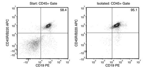
Typical EasySep™ Release Mouse Biotin Positive Selection Kit Cell Isolation Profile from Splenocytes

Figure 2. Typical EasySep™ Release Mouse Biotin Positive Selection Kit Cell Isolation Profile from Splenocytes

Starting with mouse splenocytes, the purities of the start and final isolated fractions are 58.4% and 95.1%, respectively, using a biotinylated anti-mouse CD19 antibody and EasySep™ Release Mouse Biotin Positive Selection Kit (as assessed by labeling with CD19 and CD45R/B220).

产品说明书及文档

请在《产品说明书》中查找相关支持信息和使用说明,或浏览下方更多实验方案。

Document Type
Product Name
Catalog #
Lot #
Language
Document Type
产品说明书
Catalog #
17655
Lot #
1000157902 or higher
Language
中文
Catalog #
17655
Lot #
1000157902 or higher
Language
English
Document Type
Safety Data Sheet 1
Catalog #
17655
Lot #
All
Language
English
Document Type
Safety Data Sheet 2
Catalog #
17655
Lot #
All
Language
English
Document Type
Safety Data Sheet 3
Catalog #
17655
Lot #
All
Language
English
Document Type
Safety Data Sheet 4
Catalog #
17655
Lot #
All
Language
English
Document Type
Safety Data Sheet 5
Catalog #
17655
Lot #
All
Language
English

相关材料与文献

技术资料 (10)

文献 (2)

TCF-1 promotes chromatin interactions across topologically associating domains in T cell progenitors. W. Wang et al. Nature immunology 2022 jul

Abstract

The high mobility group (HMG) transcription factor TCF-1 is essential for early T cell development. Although in vitro biochemical assays suggest that HMG proteins can serve as architectural elements in the assembly of higher-order nuclear organization,the contribution of TCF-1 on the control of three-dimensional (3D) genome structures during T cell development remains unknown. Here,we investigated the role of TCF-1 in 3D genome reconfiguration. Using gain- and loss-of-function experiments,we discovered that the co-occupancy of TCF-1 and the architectural protein CTCF altered the structure of topologically associating domains in T cell progenitors,leading to interactions between previously insulated regulatory elements and target genes at late stages of T cell development. The TCF-1-dependent gain in long-range interactions was linked to deposition of active enhancer mark H3K27ac and recruitment of the cohesin-loading factor NIPBL at active enhancers. These data indicate that TCF-1 has a role in controlling global genome organization during T cell development.
Megakaryocytic IGF1 coordinates activation and ferroptosis to safeguard hematopoietic stem cell regeneration after radiation injury Cell Communication and Signaling : CCS 2024 May

Abstract

BackgroundHematopoietic stem cell (HSC) regeneration underlies hematopoietic recovery from myelosuppression,which is a life-threatening side effect of cytotoxicity. HSC niche is profoundly disrupted after myelosuppressive injury,while if and how the niche is reshaped and regulates HSC regeneration are poorly understood.MethodsA mouse model of radiation injury-induced myelosuppression was built by exposing mice to a sublethal dose of ionizing radiation. The dynamic changes in the number,distribution and functionality of HSCs and megakaryocytes were determined by flow cytometry,immunofluorescence,colony assay and bone marrow transplantation,in combination with transcriptomic analysis. The communication between HSCs and megakaryocytes was determined using a coculture system and adoptive transfer. The signaling mechanism was investigated both in vivo and in vitro,and was consolidated using megakaryocyte-specific knockout mice and transgenic mice.ResultsMegakaryocytes become a predominant component of HSC niche and localize closer to HSCs after radiation injury. Meanwhile,transient insulin-like growth factor 1 (IGF1) hypersecretion is predominantly provoked in megakaryocytes after radiation injury,whereas HSCs regenerate paralleling megakaryocytic IGF1 hypersecretion. Mechanistically,HSCs are particularly susceptible to megakaryocytic IGF1 hypersecretion,and mTOR downstream of IGF1 signaling not only promotes activation including proliferation and mitochondrial oxidative metabolism of HSCs,but also inhibits ferritinophagy to restrict HSC ferroptosis. Consequently,the delicate coordination between proliferation,mitochondrial oxidative metabolism and ferroptosis ensures functional HSC expansion after radiation injury. Importantly,punctual IGF1 administration simultaneously promotes HSC regeneration and hematopoietic recovery after radiation injury,representing a superior therapeutic approach for myelosuppression.ConclusionsOur study identifies megakaryocytes as a last line of defense against myelosuppressive injury and megakaryocytic IGF1 as a novel niche signal safeguarding HSC regeneration.Supplementary InformationThe online version contains supplementary material available at 10.1186/s12964-024-01651-5.

更多信息

更多信息
物种 小鼠
Magnet Compatibility • EasySep™ Magnet (Catalog #18000) • “The Big Easy” EasySep™ Magnet (Catalog #18001) • EasyPlate™ Magnet (Catalog #18102) • EasyEights™ Magnet (Catalog #18103)
样本来源 其它细胞系, 脾脏, 骨髓
Selection Method Positive

质量声明:

产品仅供研究使用,不用于针对人或动物的诊断或治疗。 欲获悉更多关于STEMCELL的质控信息,请访问 STEMCELL.CN/COMPLIANCE.

Copyright © 2026 by STEMCELL Technologies. All rights reserved.

在线联系